Software & Microscope Integrated Systems

MBF Bioscience > MBF Products & Service Solutions  > Software & Microscope Integrated Systems

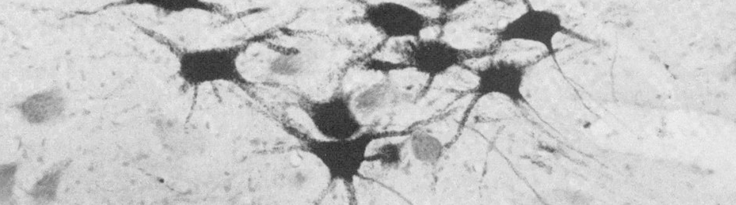
Using specimens that were collected over three decades from zoos, researchers at Humboldt University of Berlin examined facial motor control in African and Asian elephants. As described in their recent paper in Science Advances, they examined cell number, size, and position in the facial nucleus; conducted quantitative nerve tracing, and performed comparative analyses with other animals and between the two elephant types. The researchers found...

Read More

We are pleased to announce that the International Neuroinformatics Coordinating Facility (INCF) has endorsed the MBF Bioscience neuromorphological file format as a standard. The file format is used in our products for neuroscience research for important applications such as digital neuron tracing, brain mapping and stereological analyses. MBF Bioscience products, including Neurolucida, Neurolucida 360, Stereo Investigator, Vesselucida 360, Tissue Mapper and NeuroInfo use this neuromorphological file...

Read More

When we flex our thumb or point our finger, axons carry impulses from the brain to neurons in the spinal cord, which send messages to the muscles in our hands. In an important study in 1983, Jenny and Inukai at the Washington University School of Medicine reported the organizational patterns of those finger movement motoneuron columns in the primate spinal cord. Now, nearly 40 years...

Read More

Our health depends on the ability of blood vessels to deliver nutrients and remove metabolic byproducts from organs and muscle systems. But what happens to this delicately balanced process after traumatic injury? Scientists generally understand that skeletal muscles can regenerate, but little is known about how this happens at the level of our microvasculature. [caption id="attachment_7659" align="aligncenter" width="699"] Representative maps of resistance networks from feed artery...

Read More

As an unborn baby develops in the womb, its growth depends on a variety of factors, genetics among them. But sometimes a fetus doesn’t grow as much as is normally expected in relation to its gestational age. This is called intrauterine growth restriction (IUGR) or fetal growth restriction (FGR).

Babies with IUGR may develop health problems such as low resistance to infection. They may also have a hard time handling the stress of a vaginal birth. One possible cause of IUGR is that the fetus is not getting enough nutrients from the placenta.

In order to learn more about the structural differences in placentas in normal versus IUGR pregnancies, scientists at the Ludwig Maximilian University of Munich used Stereo Investigator to image tissue in both cases–finding that there are indeed quantifiable differences between the two.

One main difference is that the villi, the finger-like structures that allow nutrients and oxygen to flow from the mother to the baby, are smaller in volume in IUGR cases. Of the two types of villi present in a pregnancy, only one type—the contractile villi (the ones with muscle cells in their surrounding sheaths) were smaller. There was no difference in size between non-contractile villi in normal and IUGR placentas.

The figure shows Tukey plots of core clinical and gross anatomic data.

Read More

Our state-of-the-art software for performing unbiased stereology is getting an upgrade. Faster, stronger, and better, has been a theme for us lately, as we improve our products across the board, but one place where this is especially striking is in Stereo Investigator. Set for release this spring, the new and improved Stereo Investigator will include a new imaging engine, display engine, automatic camera alignment, automatic lens calibration, the double disector, and live video zooming.   “I’m...

Read More

Researchers at Colorado College may have identified a new neuropathological hallmark of Chronic Traumatic Encephalopathy (CTE). Their findings, published in the Journal of Comparative Neurology, describe overall dendritic atrophy across cortical neurons and greater morphological variability in CTE brains compared to controls. A neurodegenerative disorder characterized by late-onset symptoms like depression, confusion, and memory loss, CTE is caused by repeated impacts to the brain. The disease...

Read More

Scientists at Western Sydney University used Stereo Investigator and Neurolucida 360 to quantify cells in a mouse model of neuroinflammation after feeding mice two different curcumin formulations.

Some inflammation is normal in a healthy mammalian brain. But as the brain ages, processes can break down, leading to chronic neuroinflammation. This can develop into Alzheimer’s disease, dementia, and other neurodegenerative diseases.

Scientists at Prof. Gerald Muench’s lab, at Western Sydney University say that curcumin, a substance in the spice turmeric, has the potential to lower inflammation in the brain.

In two recent studies, the researchers, led by Dr. Erika Gyengesi, used Stereo Investigator and Neurolucida 360 to reconstruct and quantify glial cells in the brains of mice after feeding them two different curcumin formulations.

“MBF Bioscience’s software helped us immensely to differentiate and follow the changes caused by chronic microglia activation in various areas of the brain during aging, but also to quantify the effects of different modified curcumin products, which otherwise would have been impossible,” said Dr. Gyengesi.

In a study published February, 2020 in Scientific Reports: “Effects of a solid lipid curcumin particle formulation on chronic activation of microglia and astroglia in the GFAP-IL6 mouse model,” (Ullah et al, 2020), the researchers describe positive results after feeding GFAP-IL6 mice — a mouse model of chronic neuroinflammation — 500 ppm of Longvida®Optimised Curcumin (LC) over a course of six months.

Effect of MC on the morphological characteristics of microglial cells in the hippocampus. (A) Morphological assessment of reactive and non-reactive microglia in the hippocampus. (B–H) Microglia in the inflamed mice have significantly larger soma area, soma perimeter and processes compared with the WT mice. High dose MC significantly reduced soma area and soma perimeter compared with GFAP-IL6 mice. However, the same high dose MC significantly increased the number of nodes compared with the GFAP-Il6 mice. It has no effect on the convex area, convex perimeter, dendritic length and number of processes. Significance = *p < 0.05, **p < 0.001, ***p < 0.0001, ****p < 0.0001.

Stereological analysis of the mouse brains revealed lower levels of activated microglia in the hippocampus (26 percent less) and in the cerebellum (48 percent less) in GFAP-IL6 mice that were fed the curcumin diet, compared to GFAP-IL6 mice fed a normal diet. They also quantified astrocytes — another cell type activated in response to neuroinflammation, finding decreased levels in the hippocampus (30 percent less). TSPO+ cells — another marker of brain inflammation, decreased as well (by 24 percent in the hippocampus and 31 percent in the cerebellum) in the experimental mice compared to controls.

Read More

Researchers Quantify Improvement in Heart Vasculature with Vesselucida 360 and Vesselucida Explorer Cells need oxygen to survive, but during a heart attack, blood flow is restricted and cardiac cells can’t get the oxygen they need to stay alive. A new therapy, developed by researchers at the Coulombe Lab at Brown University may be able to provide the heart with the support it needs to recover after...

Read More

For Immediate Release: Williston, VT (February 04, 2020) — MBF Bioscience’s revolutionary light sheet microscope system, ClearScope, sets a new standard for microscopic imaging. The new decade is poised to bring about incredible scientific innovations, and MBF Bioscience is leading the charge in 2020 with the creation of the “light sheet theta microscope” system, ClearScope. MBF Bioscience secured exclusive license from Columbia University to develop the light...

Read More