
3D Reconstructions of Neurons Reveal More Branching in Sedentary Rats
Scientists discovered that inactivity makes brain cells grow, but not in a good way. In a study published in the Journal of Comparative Neurology, researchers found more neuronal branching in sedentary rats compared to active rats. The growth occurred in a region of the brain that controls blood pressure, leading the scientists to hypothesize that these changes may be part of the reason inactivity is linked to an increased risk of heart disease.
Using Neurolucida to reconstruct neurons in 3D, the scientists at Dr. Patrick Mueller’s lab at Wayne State University School of Medicine, in Detroit, saw structural differences between the brains of active and inactive rats.
Focusing on the rostral ventrolateral medulla (RVLM) – an area that controls several critical biological processes that rats as well as humans do unconsciously, like swallowing, breathing, and regulating blood pressure, the scientists saw longer dendrites, more dendritic branching, and more intersections with other neurons in sedentary rats.
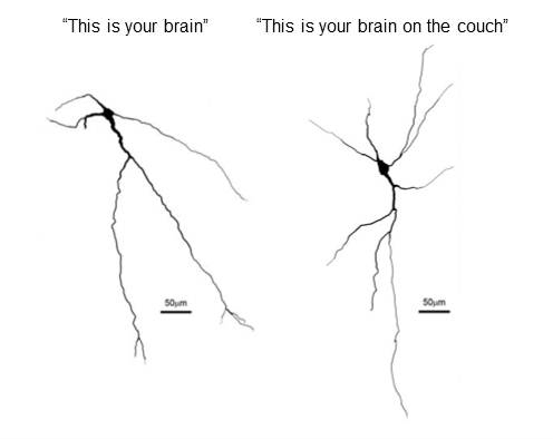
Left: A neuron from the brain of a rat that exercised for two hours each day.
Right: A neuron from the brain of a sedentary rat. Scientists saw greater branching in inactive versus active rats. (Image courtesy of Dr. Patrick Mueller)
The rats studied were divided into two groups, six of the animals ran in an exercise wheel for about two hours per day over the course of about three months, while the remaining five were housed in cages without wheels and experienced very little activity.
After eleven or twelve weeks, the researchers injected the rats with a tracer to mark specific neurons in the rostral ventrolateral medulla. To reconstruct the neurons in Neurolucida, they used images of immunostained sections of both sets of rats’ brains.
In their paper, the authors say the robust branching they saw in inactive rats may make RVLM neurons more sensitive to stimuli and increase excitability of the sympathetic nervous system, which is known to play a role in high blood pressure and heart disease.
Mischel, N. A., Llewellyn-Smith, I. J., & Mueller, P. J. (2013). Physical (in)activity dependent structural plasticity in bulbospinal catecholaminergic neurons of rat rostral ventrolateral medulla. Journal of Comparative Neurology, n/a-n/a. Doi: 10.1002/cne.23464.