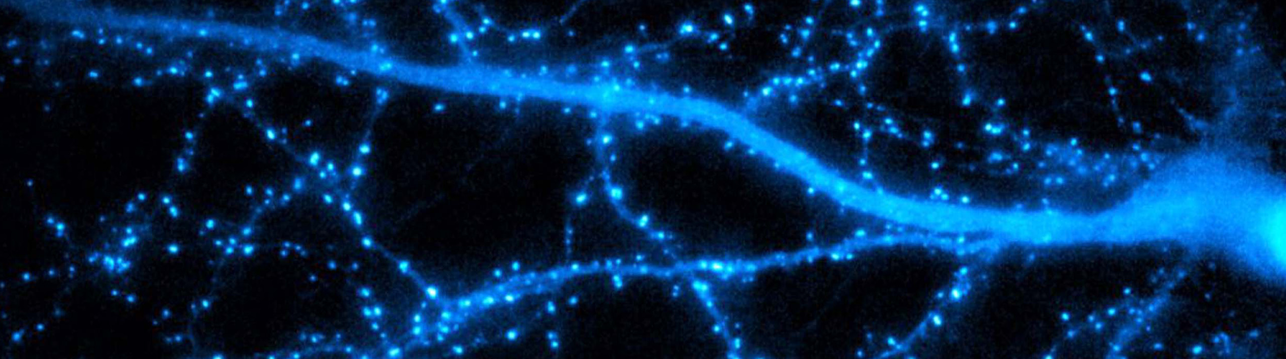
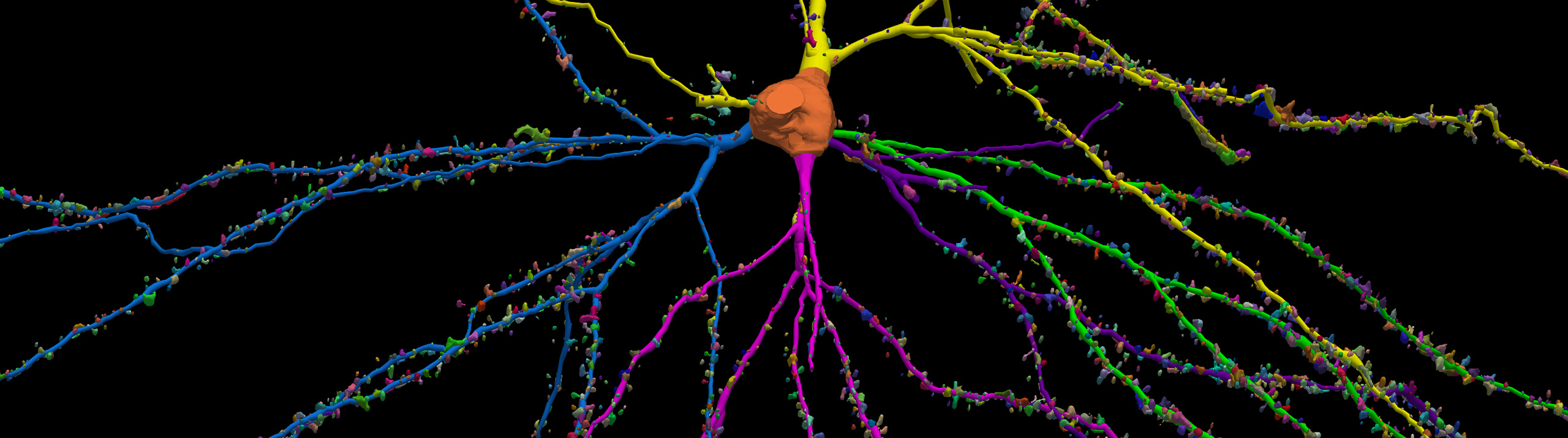
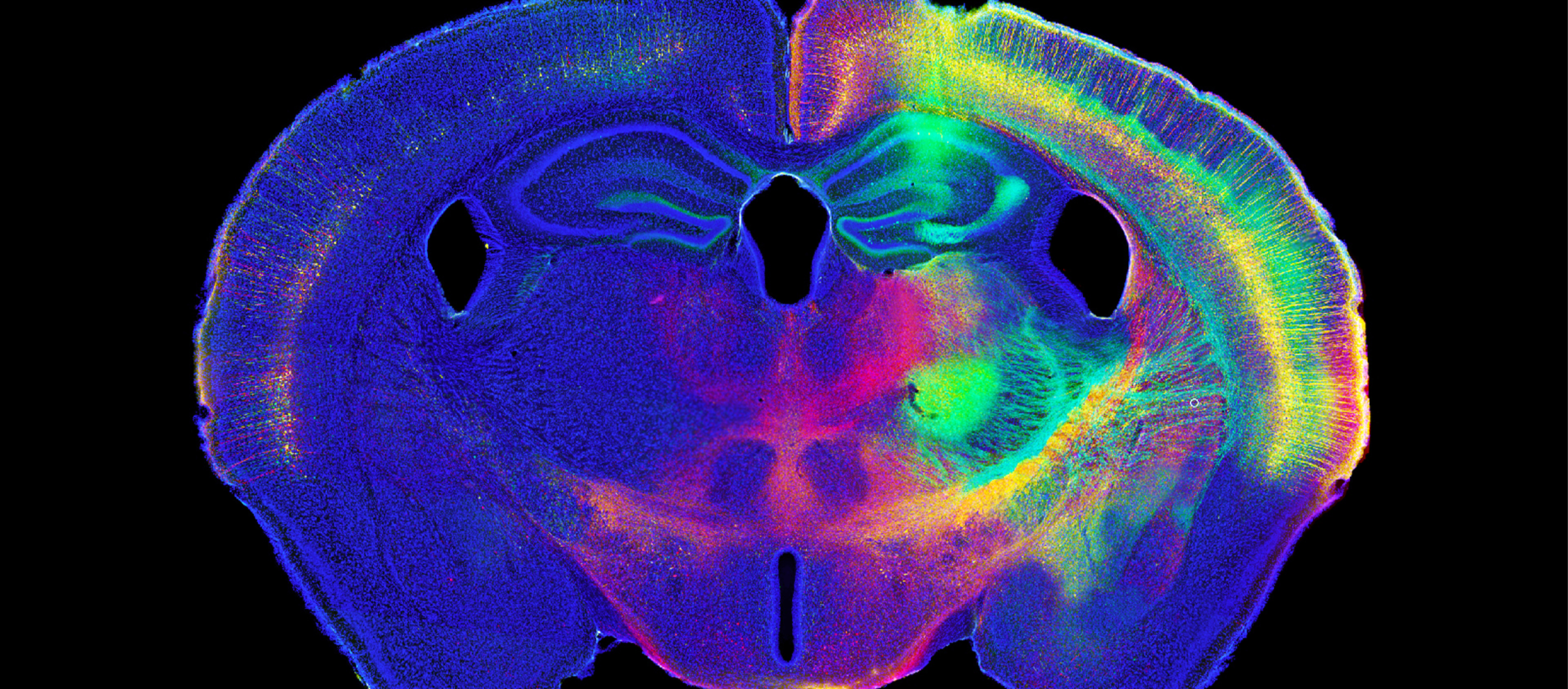
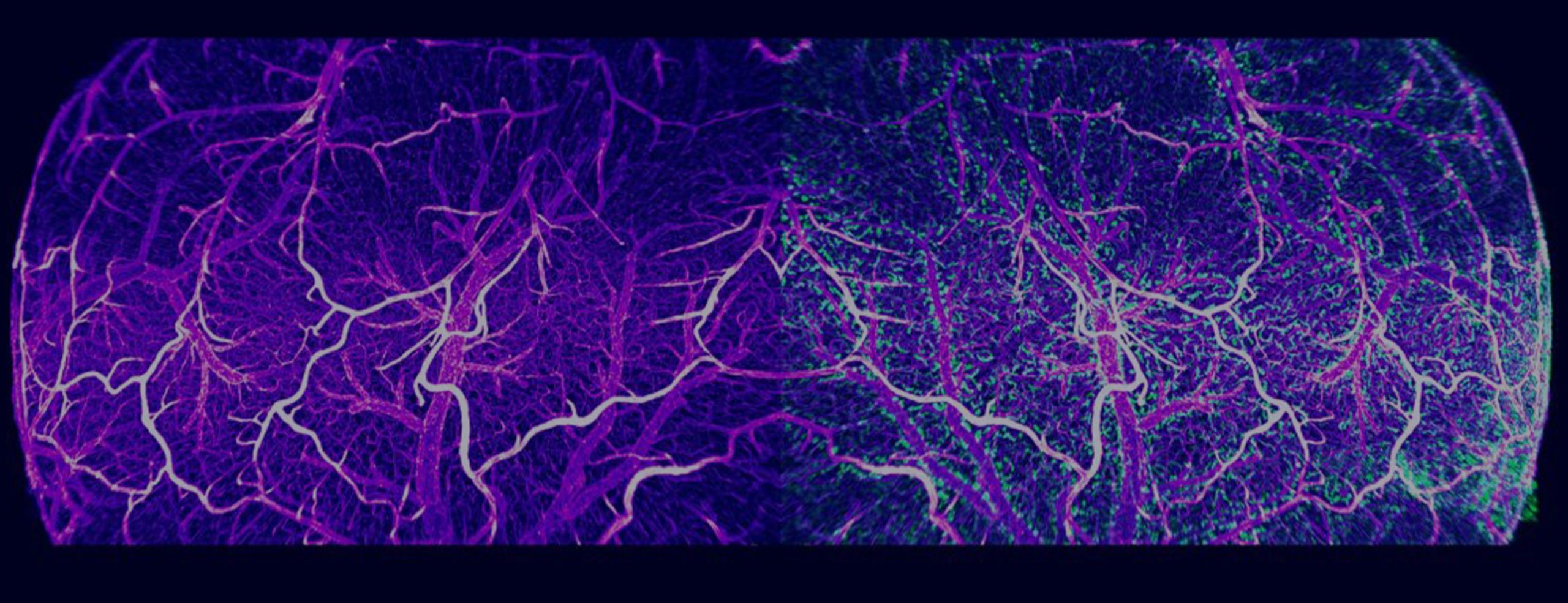
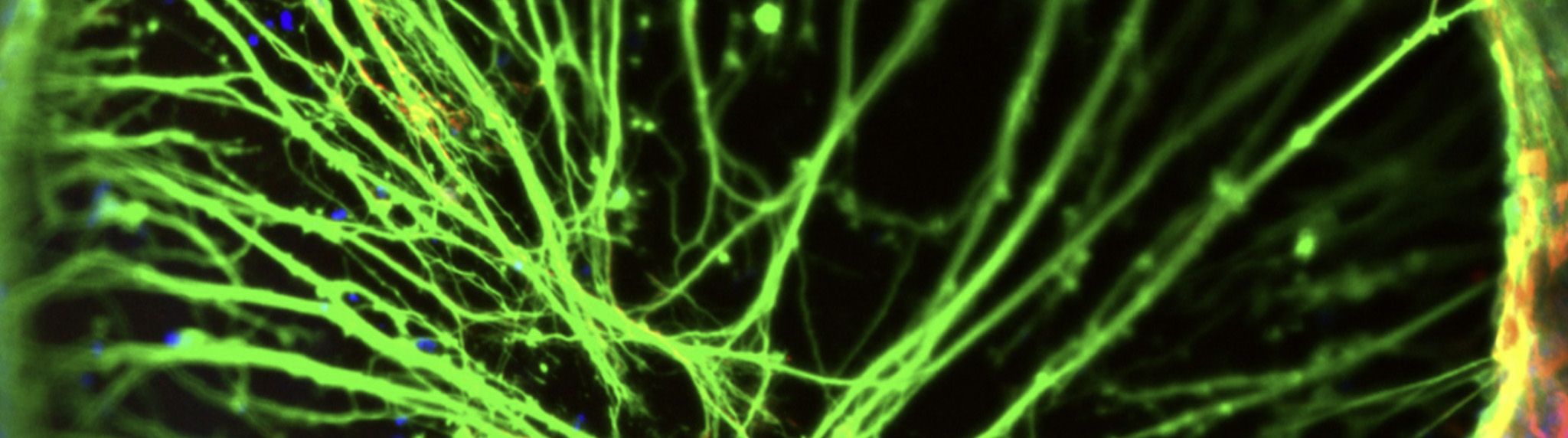
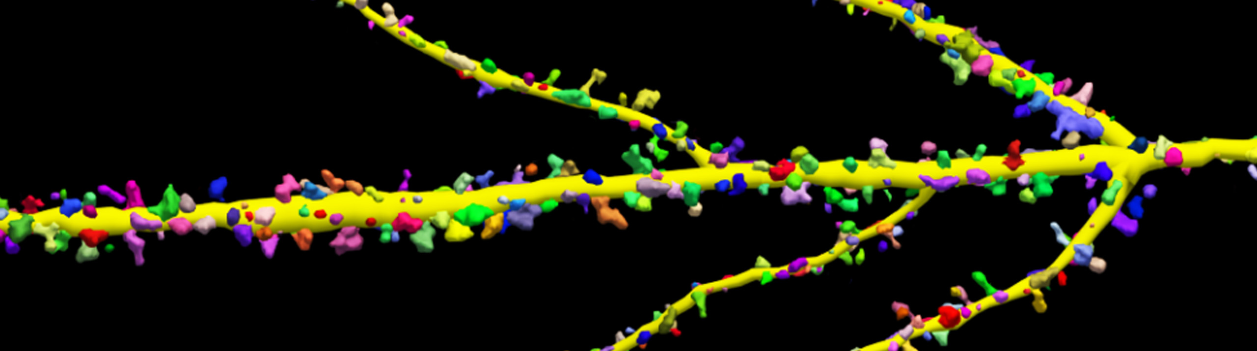
Software Applications For Quantitive Analysis

MBF Bioscience > MBF Products & Service Solutions  > Software Applications For Quantitive Analysis

The Image Volume Fractionator probe, available in Stereo Investigator - Cleared Tissue Edition, is facilitating huge efficiency gains for quantifying the number of cells. At Dr. Patrick R. Hof’s lab at the Icahn School of Medicine at Mount Sinai, researchers imaged the cerebral cortex using light-sheet fluorescence microscopy and quantified the number of neurons, including those that express proteins involved in Alzheimer’s disease and schizophrenia, using...

Read More

After decades of identifying brain cells subjectively, researchers can now make use of a standardized classification system for identifying pyramidal cells—the most common type of cells in the neocortex. Scientists at the Blue Brain Project developed the system using mathematics that identify the properties of shapes that stay constant under continuous transformation. This new method of classification gives researchers the ability to begin building a...

Read More

We are pleased to announce that the International Neuroinformatics Coordinating Facility (INCF) has endorsed the MBF Bioscience neuromorphological file format as a standard. The file format is used in our products for neuroscience research for important applications such as digital neuron tracing, brain mapping and stereological analyses. MBF Bioscience products, including Neurolucida, Neurolucida 360, Stereo Investigator, Vesselucida 360, Tissue Mapper and NeuroInfo use this neuromorphological file...

Read More
High Resolution scan of brain

Researchers studying structure and function in rat brain can now use NeuroInfo to analyze and register their brain volumes to the Waxholm Rat Brain Atlas Version 4—an open access volumetric atlas of the Sprague Dawley rat brain. “NeuroInfo already includes extensive analysis capabilities for mouse brain research by standardizing measurements on brain volumes to the Allen Mouse Brain Atlas. The inclusion of a rat atlas in...

Read More

Our health depends on the ability of blood vessels to deliver nutrients and remove metabolic byproducts from organs and muscle systems. But what happens to this delicately balanced process after traumatic injury? Scientists generally understand that skeletal muscles can regenerate, but little is known about how this happens at the level of our microvasculature. [caption id="attachment_7659" align="aligncenter" width="699"] Representative maps of resistance networks from feed artery...

Read More

The heart has a “little brain.” It’s a network of neurons known as the intrinsic cardiac nervous system (ICNS), and it plays a key role in regulating cardiac activity. Building on previous research (Achanta et al., 2020), which resulted in a 3D map of the rat ICNS, a new study by a team of scientists from the University of Central Florida, Thomas Jefferson University, the University...

Read More

MBF Bioscience is now leveraging how neurons learn in order to improve neuroscience research using microscopy. By incorporating artificial neural networks into MBF Bioscience software, we’re equipping neuroscientists with tools that characterize neuronal populations with unprecedented accuracy and anatomic specificity through entire brain volumes. In the webinar titled, “Improved detection of c-fos labeled and pyramidal neurons using deep machine learning in NeuroInfo,” Dr. Gerfen, joined by...

Read More

For Immediate Release Williston, VT (February 11th, 2021)—MicroDynamixsoftware version 2021 is now available. This state-of-the-art dendritic-spine analysis application enables users to visualize and quantify spine morphology over time using powerful algorithms and detection methods. Developed with input and support from the National Institute of Mental Health (NIMH), the new version of MicroDynamix software makes it easier than ever for neuroscientists to obtain accurate 4D dynamic analyses...

Read More

For Immediate Release   Williston, VT (December 10, 2020) —NeuroInfo®is the most advanced software for mapping brain sections, 3D volumes and cell populations into a standardized atlas reference space.  Optimized to enable researchers to work far more efficiently, the new NeuroInfo update means faster, more efficient measurements and mapping workflows.  The new version of NeuroInfo includes:  Identification and delineation of brain regions in experimental mouse-brain sections  Automatic cell detection using deep learning to recognize and count specific neuronal subtypes  Performance optimizations that...

Read More

Scientists at Western Sydney University used Stereo Investigator and Neurolucida 360 to quantify cells in a mouse model of neuroinflammation after feeding mice two different curcumin formulations.

Some inflammation is normal in a healthy mammalian brain. But as the brain ages, processes can break down, leading to chronic neuroinflammation. This can develop into Alzheimer’s disease, dementia, and other neurodegenerative diseases.

Scientists at Prof. Gerald Muench’s lab, at Western Sydney University say that curcumin, a substance in the spice turmeric, has the potential to lower inflammation in the brain.

In two recent studies, the researchers, led by Dr. Erika Gyengesi, used Stereo Investigator and Neurolucida 360 to reconstruct and quantify glial cells in the brains of mice after feeding them two different curcumin formulations.

“MBF Bioscience’s software helped us immensely to differentiate and follow the changes caused by chronic microglia activation in various areas of the brain during aging, but also to quantify the effects of different modified curcumin products, which otherwise would have been impossible,” said Dr. Gyengesi.

In a study published February, 2020 in Scientific Reports: “Effects of a solid lipid curcumin particle formulation on chronic activation of microglia and astroglia in the GFAP-IL6 mouse model,” (Ullah et al, 2020), the researchers describe positive results after feeding GFAP-IL6 mice — a mouse model of chronic neuroinflammation — 500 ppm of Longvida®Optimised Curcumin (LC) over a course of six months.

Effect of MC on the morphological characteristics of microglial cells in the hippocampus. (A) Morphological assessment of reactive and non-reactive microglia in the hippocampus. (B–H) Microglia in the inflamed mice have significantly larger soma area, soma perimeter and processes compared with the WT mice. High dose MC significantly reduced soma area and soma perimeter compared with GFAP-IL6 mice. However, the same high dose MC significantly increased the number of nodes compared with the GFAP-Il6 mice. It has no effect on the convex area, convex perimeter, dendritic length and number of processes. Significance = *p < 0.05, **p < 0.001, ***p < 0.0001, ****p < 0.0001.

Stereological analysis of the mouse brains revealed lower levels of activated microglia in the hippocampus (26 percent less) and in the cerebellum (48 percent less) in GFAP-IL6 mice that were fed the curcumin diet, compared to GFAP-IL6 mice fed a normal diet. They also quantified astrocytes — another cell type activated in response to neuroinflammation, finding decreased levels in the hippocampus (30 percent less). TSPO+ cells — another marker of brain inflammation, decreased as well (by 24 percent in the hippocampus and 31 percent in the cerebellum) in the experimental mice compared to controls.

Read More