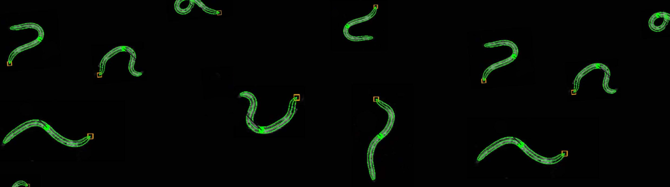
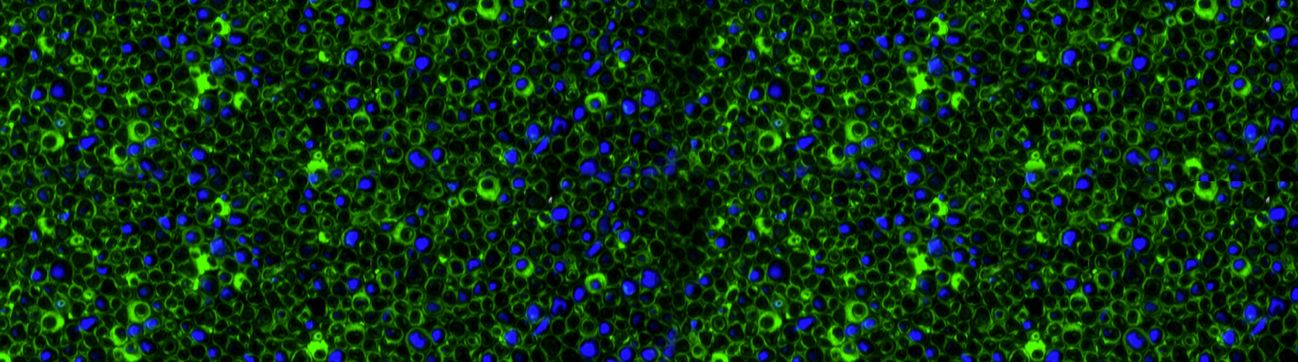
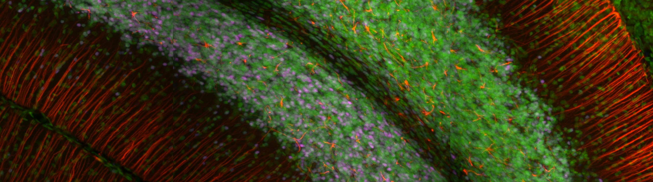
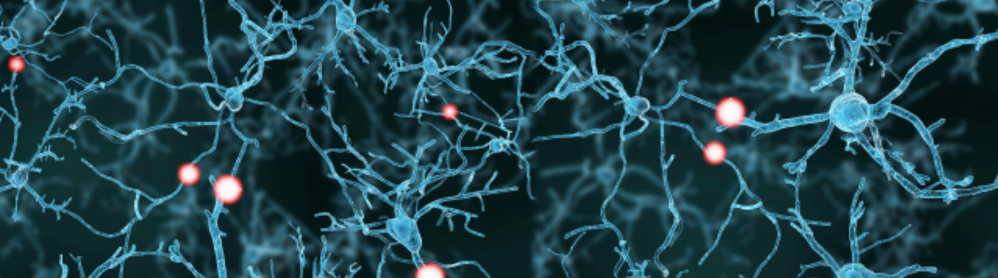
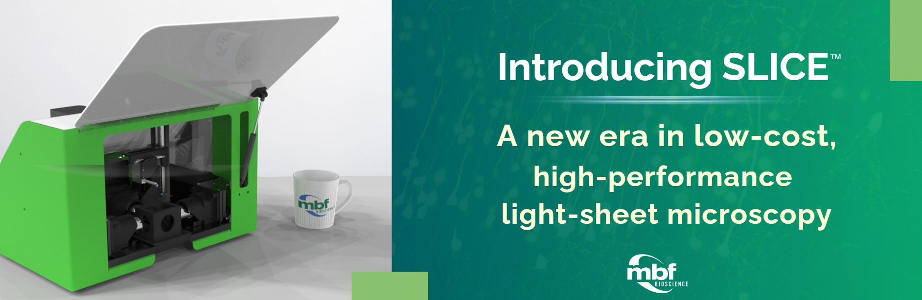
Scientific Applications & Use Cases

MBF Bioscience > Scientific Applications & Use Cases

ScanImage®software from MBF Bioscience, along with the accompanying vDAQTM acquisition and control card with analog to digital, digital to analog, breakout board, is built to control many combinations of hardware in order to carry out in-vivo imaging on a cellular scale. This makes it possible to observe the neural activities, such as those indicated by calcium concentration or voltage changes, of specific neuronal types...

Read More

Using specimens that were collected over three decades from zoos, researchers at Humboldt University of Berlin examined facial motor control in African and Asian elephants. As described in their recent paper in Science Advances, they examined cell number, size, and position in the facial nucleus; conducted quantitative nerve tracing, and performed comparative analyses with other animals and between the two elephant types. The researchers found...

Read More

The Image Volume Fractionator probe, available in Stereo Investigator - Cleared Tissue Edition, is facilitating huge efficiency gains for quantifying the number of cells. At Dr. Patrick R. Hof’s lab at the Icahn School of Medicine at Mount Sinai, researchers imaged the cerebral cortex using light-sheet fluorescence microscopy and quantified the number of neurons, including those that express proteins involved in Alzheimer’s disease and schizophrenia, using...

Read More

Humans and animal species can move in controlled motion sequences because of a delicate balance in the signaling of certain neurons in the area of the brain called the striatum. In this brain region, some neurons tell muscles to move, while others tell muscles to hold steady. In the brains of patients with Parkinson’s disease, more of the neurons that signal movement are activated resulting...

Read More

Obesity has reached epidemic levels worldwide. It is a major cause of deadly illnesses, such as diabetes, cardiovascular disease, and some cancers, and has been linked to reduced quality of life and poor mental health outcomes. Efforts to combat this epidemic have so far failed to have an appreciable impact. Seeking novel targets with the potential to move the needle on obesity-linked cardiometabolic disorders, a research...

Read More

Most neuroscientists are familiar with the saying “cells that fire together wire together,” which is often used to summarize the Hebbian theory of synaptic plasticity—first put forth in Donald Hebb’s book The Organization of Behavior in 1949. The theory describes how coincident activity between pre- and post-synaptic cells can shape synaptic strength. Verified decades later, the theory has since become accepted within the neuroscientific community. However,...

Read More

What if, instead of trying to zoom in on the nanoscale structures of the brain, we made them bigger? Dr. Edward Boyden and his team at MIT are doing just that. The process, known as expansion microscopy (ExM), physically enlarges brain tissue so that even extremely small molecular structures are viewable on a conventional light microscope. In his 2016 TED Talk, Dr. Boyden says: “Can we make...

Read More

As an unborn baby develops in the womb, its growth depends on a variety of factors, genetics among them. But sometimes a fetus doesn’t grow as much as is normally expected in relation to its gestational age. This is called intrauterine growth restriction (IUGR) or fetal growth restriction (FGR).

Babies with IUGR may develop health problems such as low resistance to infection. They may also have a hard time handling the stress of a vaginal birth. One possible cause of IUGR is that the fetus is not getting enough nutrients from the placenta.

In order to learn more about the structural differences in placentas in normal versus IUGR pregnancies, scientists at the Ludwig Maximilian University of Munich used Stereo Investigator to image tissue in both cases–finding that there are indeed quantifiable differences between the two.

One main difference is that the villi, the finger-like structures that allow nutrients and oxygen to flow from the mother to the baby, are smaller in volume in IUGR cases. Of the two types of villi present in a pregnancy, only one type—the contractile villi (the ones with muscle cells in their surrounding sheaths) were smaller. There was no difference in size between non-contractile villi in normal and IUGR placentas.

The figure shows Tukey plots of core clinical and gross anatomic data.

Read More

MBF Bioscience is now leveraging how neurons learn in order to improve neuroscience research using microscopy. By incorporating artificial neural networks into MBF Bioscience software, we’re equipping neuroscientists with tools that characterize neuronal populations with unprecedented accuracy and anatomic specificity through entire brain volumes. In the webinar titled, “Improved detection of c-fos labeled and pyramidal neurons using deep machine learning in NeuroInfo,” Dr. Gerfen, joined by...

Read More

Our state-of-the-art software for performing unbiased stereology is getting an upgrade. Faster, stronger, and better, has been a theme for us lately, as we improve our products across the board, but one place where this is especially striking is in Stereo Investigator. Set for release this spring, the new and improved Stereo Investigator will include a new imaging engine, display engine, automatic camera alignment, automatic lens calibration, the double disector, and live video zooming.   “I’m...

Read More