
Neurolucida helps to reveal new findings in a follow-up to a 40 year old study
When we flex our thumb or point our finger, axons carry impulses from the brain to neurons in the spinal cord, which send messages to the muscles in our hands. In an important study in 1983, Jenny and Inukai at the Washington University School of Medicine reported the organizational patterns of those finger movement motoneuron columns in the primate spinal cord. Now, nearly 40 years later, Paul Cheney at the University of Kansas Medical Center and Arthur B. Jenny, an author from the original study, are using Neurolucida to further analyze these neurons, and they have made some noteworthy new findings.
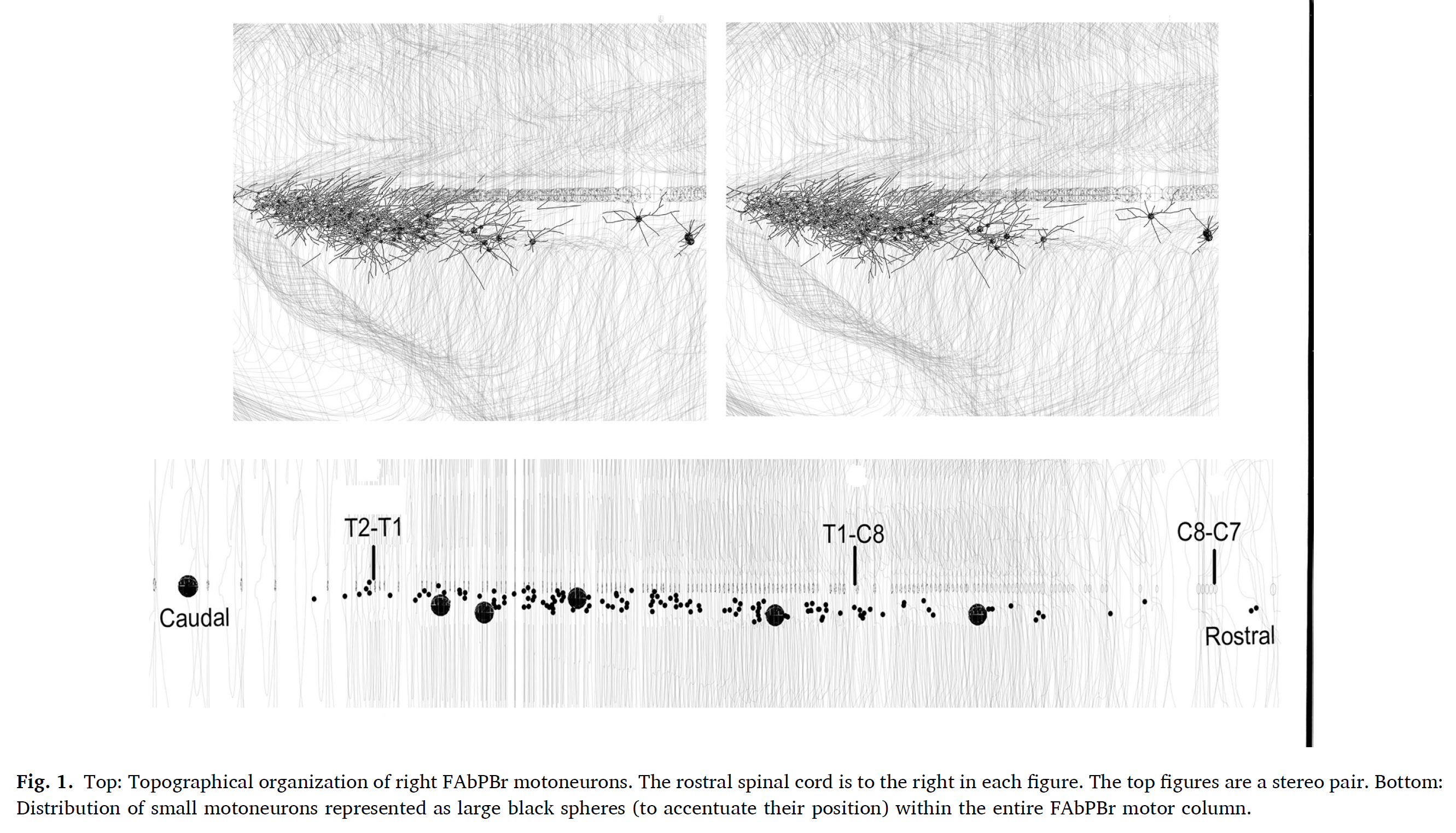
According to Dr. Jenny, “the 3D capabilities of Neurolucida has made it possible to better understand the distribution of small motoneurons within the motor column, and the patterns of motoneuron dendrites within the spinal cord.” The researchers are interested in comparing the patterns of motoneuron dendritic trees with the known patterns of brain-spinal cord terminal connections.
“We believe a better understanding of the motor column dendritic trees relative to descending supra-spinal inputs can be used to guide rehabilitation efforts in people recovering from stroke or spinal cord injury.”(Jenny, Cheney, 2022)
© Elsevier B.V. All rights reserved.
Over the last four decades, advances in technology such as injections of anatomical tracers into motoneurons have provided a clearer picture of the entire dendritic tree. Dendritic trees appear to extend outward from the cell body in mostly linear and radial directions before branching. In the current research, only the proximal parts of the dendritic trees were labeled with tracer, but the patterns of the proximal dendrites were similar to the patterns seen with intracellular injections of tracer. The researchers considered the direction of dendrites seen in their material to be a reasonable estimate for the direction of the more distal unlabeled dendrites.
In the current study, the researchers used Neurolucida to digitally reconstruct the neurons from the original microscopic slides from 1983 to perform a detailed 3D analysis of dendritic tree patterns. While they observed dendrites radiating in all directions, this new analysis revealed a preference for two specific directions (either toward the gray matter at the base of the dorsal horn or to the gray matter of the medial ventral horn) suggesting the possibility that motoneuron dendritic trees extend toward functional terminal regions.
The authors plan to use Neurolucida to continue re-examining the motoneuron columns analyzed in the original 1983 study, with the ultimate goal of merging their data with data from other investigations into a 3D neuron database.
Jenny AB, Cheney PD. Monkey flexor and abductor pollicis brevis motoneuron pools: Proximal dendritic trees and small motoneurons. Neuroscience Letters. Vol. 769, 2022 Jan 19. doi: 10.1016/j.neulet.2021.136429
Jenny AB, Inukai J. Principles of Motor Organization of the Monkey Cervical Spinal Cord. Journal of Neurosci. Vol. 3, No. 3, pp. 567-575, 1983 Mar. https://www.jneurosci.org/content/jneuro/3/3/567.full.pdf