Software & Microscope Integrated Systems

MBF Bioscience >  Blog > MBF Products & Service Solutions  > Software & Microscope Integrated Systems (Page 2)

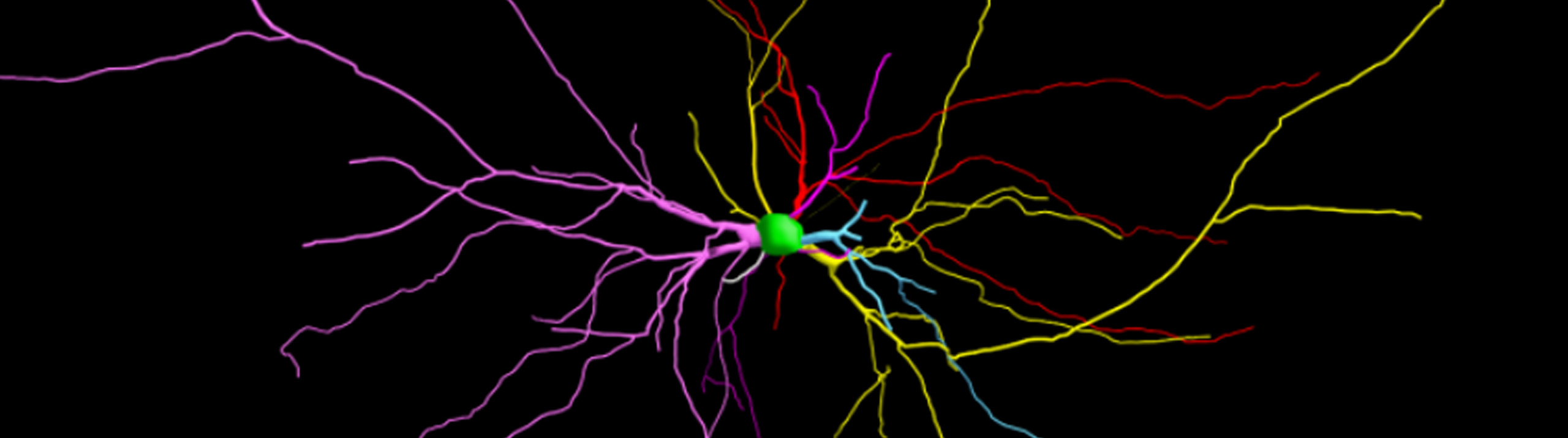
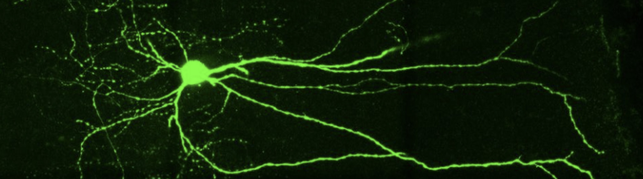
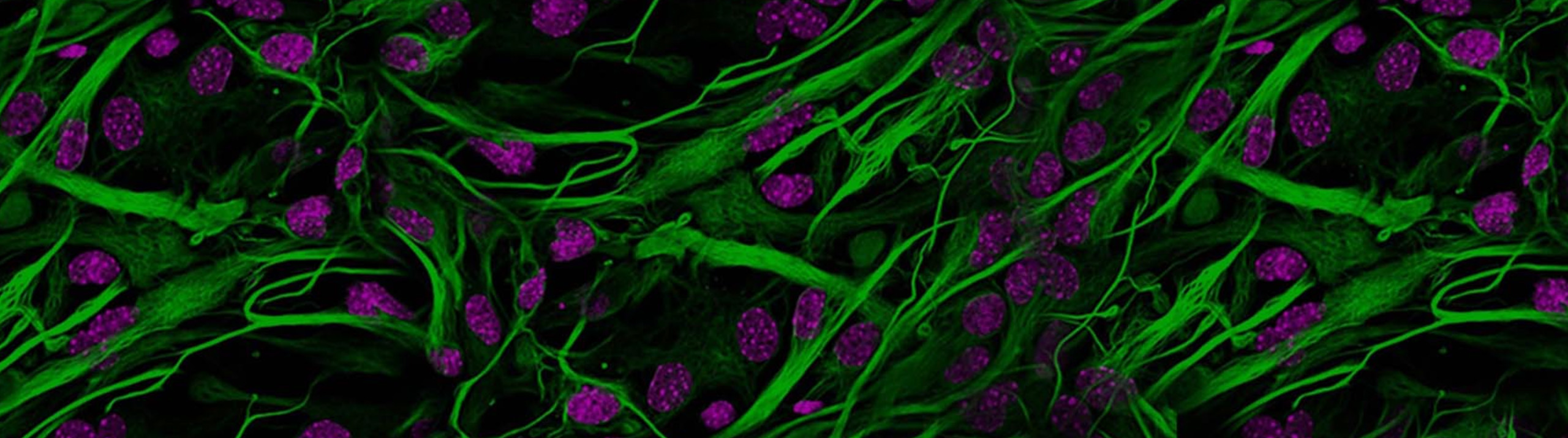
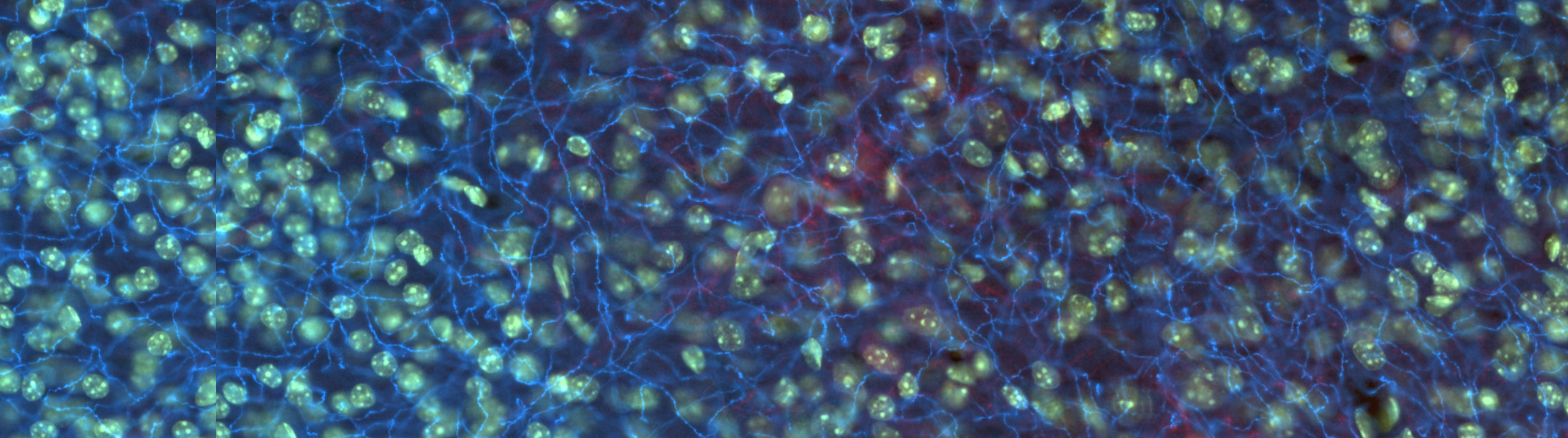
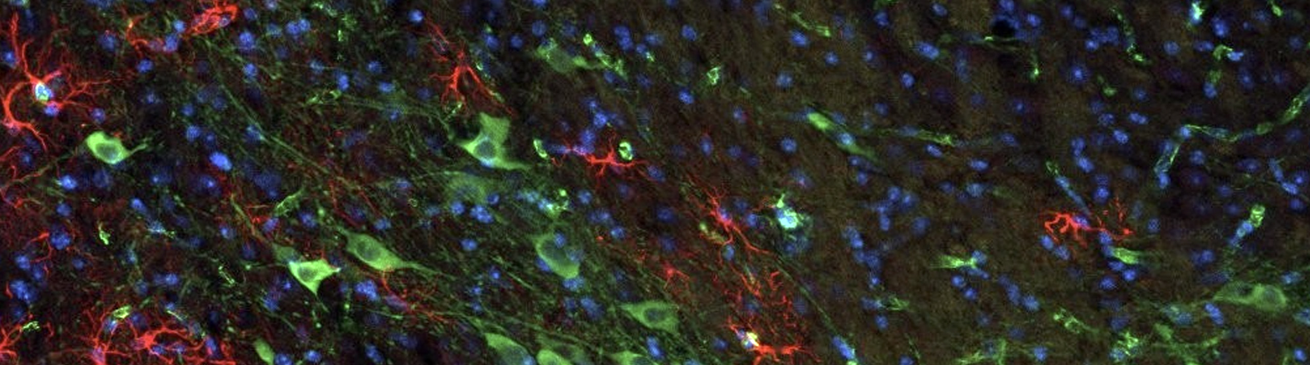
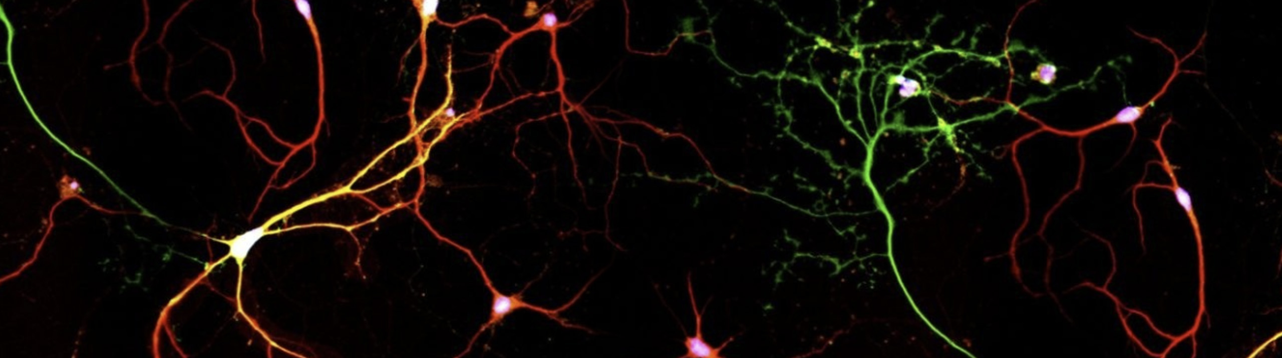
Neurolucida and Neurolucida Explorer Used for 3D Reconstruction and Quantitative Analysis Researchers used Neurolucida to reconstruct a newly discovered type of neuron found only in the human brain, according to a study published in the journal Nature Neuroscience. Known as “rosehip” neurons because of the way they resemble a rose after its petals have fallen off, these cells feature compact, bushy axonal arborizations.   Found in the first...

Read More

[caption id="attachment_6839" align="aligncenter" width="632"] Image Courtesy: Bob Jacobs, Ph.D. , Colorado College[/caption]   With the release of its new version on November 28, NeuroMorpho.org adds 9,987 new images to its archive, bringing its impressive collection of digitally reconstructed neurons to 80,012.   Scientists used MBF Bioscience’s software, Neurolucida and Neurolucida 360, to reconstruct the majority of these cells. In fact, 64 times more neurons were reconstructed with MBF Bioscience...

Read More

[caption id="attachment_6830" align="aligncenter" width="632"] An experimental coronal mouse brain section automatically aligned to the Allen Mouse Brain Reference Atlas[/caption]   Analyzing cellular populations within specific anatomies in brain images requires expertise in both neuroanatomy and cellular identification. This typically involves a scientist comparing experimental images with a reference atlas and manually delineating anatomical regions and marking cell populations within. NeuroInfo®, a revolutionary new technology from MBF Bioscience,...

Read More

  DNA damage occurs in human cells at a constant rate. These cells are usually able to repair themselves, but sometimes deficiencies in certain genes cause the repair process to shut down. When damaged DNA isn't fixed, mutations can occur that cause accelerated aging or cancerous tumors to form (Hoeijmakers, 2009). Scientists at Erasmus University Medical Center in Rotterdam have found a way to slow down...

Read More

Neuroscientists can now analyze the size and complexity of neurons and collect unbiased stereology data with greater speed and efficiency We are happy to announce the release of Neurolucida and Stereo Investigator version 2017. This version features a completely revamped user interface that’s intuitive and easy to navigate.   “Neurolucida and Stereo Investigator version 2017 are completely redesigned to improve the user experience and increase productivity,” said Jack...

Read More

Astrocytes (GFAP) in the dentate gyrus of a mouse hippocampus. Image courtesy of Dr. Ahmad Salehi, Stanford University.   It is well known that physical exercise eases the symptoms of neurodegenerative disorders like Alzheimer’s disease and helps to prevent their onset. Researchers at Stanford University are working on figuring out how it happens.   In their study, published in the journal Brain Structure and Function, scientists in Dr....

Read More

[caption id="attachment_6526" align="aligncenter" width="800"] Representative images of Iba-1+ microglia in the postnatal day 10 rat hippocampus. Image courtesy of Anna Klintsova, PhD.[/caption] Children born with fetal alcohol spectrum disorders face a range of physical and cognitive impairments including long-term deficits in learning, behavior, and immune function. In a paper published in Neuroscience, Dr. Anna Klintsova and her lab at the University of Delaware report that activation...

Read More

The optical fractionator probe was used to quantify the number of neurons and glia in the dentate gyrus   Doctors have used lithium to treat patients with bipolar disorder since the 1970s. Known for its efficacy in stabilizing patients' moods by regulating manic episodes, lithium is also associated with a decreased risk of suicide. But while this naturally occurring element is the most widely prescribed medication for...

Read More

[caption id="attachment_6403" align="aligncenter" width="632"] This figure illustrates the separate and combined effects of acute stress and fear conditioning/extinction on dendritic morphology of pyramidal neurons in the infralimbic region of medial prefrontal cortex. Each neuron shown is a composite made up of apical (blue) and basilar (orange) arbor near the mean of the group. The apical and basilar arbors of each composite are from different neurons....

Read More

[caption id="attachment_6326" align="aligncenter" width="600"] An immunostained image of myelin basic protein in the cerebella of a mouse brain with an iron-sufficient diet compared with the brain of a mouse exposed to alcohol and fed an iron-insufficient diet. It shows the reduced cerebellar size due to the ID-alcohol combination. Green is MBP immunostain, blue is DAPI for nuclei. Image courtesy of Susan Smith, PhD.[/caption]   If a pregnant...

Read More