
Differences Associated with Fetal Growth Restriction are Found Using Stereo Investigator
As an unborn baby develops in the womb, its growth depends on a variety of factors, genetics among them. But sometimes a fetus doesn’t grow as much as is normally expected in relation to its gestational age. This is called intrauterine growth restriction (IUGR) or fetal growth restriction (FGR).
Babies with IUGR may develop health problems such as low resistance to infection. They may also have a hard time handling the stress of a vaginal birth. One possible cause of IUGR is that the fetus is not getting enough nutrients from the placenta.
In order to learn more about the structural differences in placentas in normal versus IUGR pregnancies, scientists at the Ludwig Maximilian University of Munich used Stereo Investigator to image tissue in both cases–finding that there are indeed quantifiable differences between the two.
One main difference is that the villi, the finger-like structures that allow nutrients and oxygen to flow from the mother to the baby, are smaller in volume in IUGR cases. Of the two types of villi present in a pregnancy, only one type—the contractile villi (the ones with muscle cells in their surrounding sheaths) were smaller. There was no difference in size between non-contractile villi in normal and IUGR placentas.
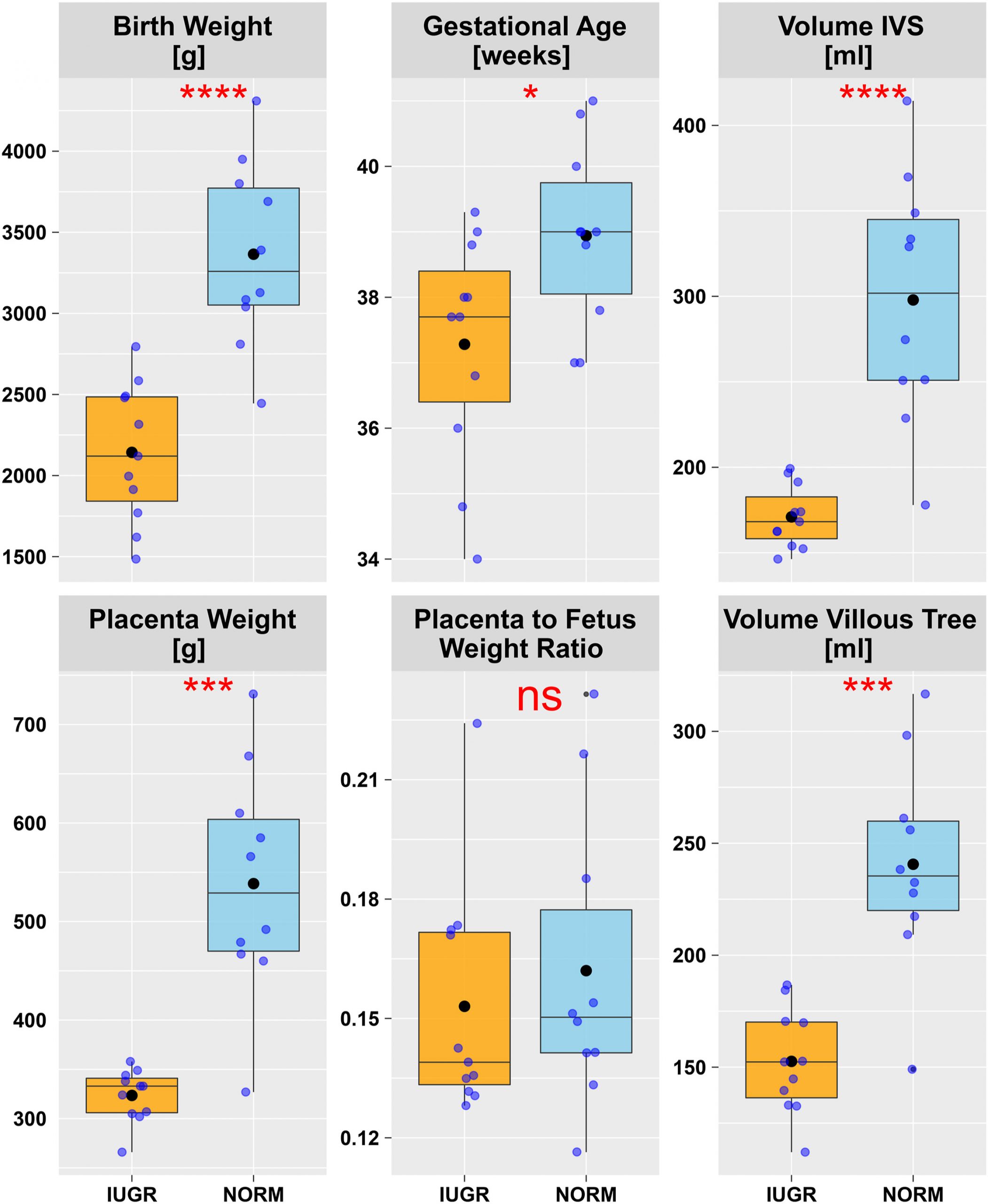
The figure shows Tukey plots of core clinical and gross anatomic data.
IUGRHowever, in both types of villi in IUGR placentas, the researchers observed reduced blood vessel volume, longer diffusion distances (distance from fetal to maternal blood), and less branching.
To achieve these results, the researchers extracted six tissue samples from 21 placentas (10 normal, 11 IUGR), and used an MBF Bioscience system comprising microscope, motorized stage, camera, z-encoder, and Stereo Investigator to image samples.
“This combination of software and hardware in the Stereo Investigator system makes it possible to image the tissue sections using systematic random sampling and perform unbiased stereology probes to estimate the percent by volume of different components of the placenta, the amount of branching of the villi, and the diffusion distance. The system allows for unbiased estimates in an efficient manner,” says MBF Bioscience Staff Scientist Dr. Dan Peruzzi.
In their study, the Munich researchers combined three Stereo Investigator probes, including the area fraction fractionator probe (used to estimate percentage by volume) and the probe used to measure diffusion distance, in a novel way.
Citation:
Barapatre, N., Kampfer, C., Henschen, S., Schmitz, C., Edler von Koch, F., Frank, H.G., Growth restricted placentas show severely reduced volume of villous components with perivascular myofibroblasts. Placenta, (109) 2021. https://doi.org/10.1016/j.placenta.2021.04.006