Unprecedented study reveals structure-function adaptations in the facial nucleus of elephants
Using specimens that were collected over three decades from zoos, researchers at Humboldt University of Berlin examined facial motor control in African and Asian elephants. As described in their recent paper in Science Advances, they examined cell number, size, and position in the facial nucleus; conducted quantitative nerve tracing, and performed comparative analyses with other animals and between the two elephant types. The researchers found that the facial nucleus in elephants is much larger than in most other mammals and that it is both larger and more complex in the African elephants than in the Asian elephants in their study. Their results suggest that elephant brains exhibit neural adaptations related to facial morphology and dexterity, and overall body size.
The facial nucleus, present in vertebrate animals, is a group of neurons in the brainstem that receives instructions from neurons in the cortex to direct movement of the muscles of the face. For this paper, the authors characterized the facial nucleus of two similar species, African and Asian elephants, with muscular, dexterous trunks. These species’ faces share many similarities, but have distinctly different ear size and trunk morphology—areas controlled by the facial nucleus. The authors used varied methods, matched to the available elephant material, including Nissl staining, cell counting, axonal osmium tetroxide stains, somata drawings, cell fiber counting, and nerve tracing to examine the facial nucleus in specimens from African (n=4) and Asian elephants (n=4 elephants). Stereo Investigator® software was used to acquire images of thin sections, conduct stereological procedures, and measure cell size and axon diameter.
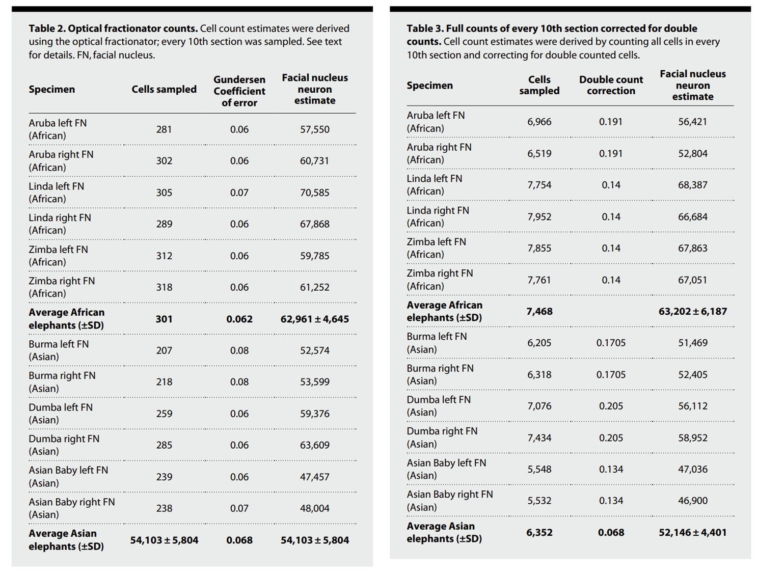
Two methods were used to quantify cell populations in the elephant facial nucleus, an unbiased stereology approach and a model-based stereology strategy that consisted of complete counts of cell pieces in every tenth section were used. Using unbiased stereology, the researchers counted ~200-300 cells per specimen with the Stereo Investigator optical fractionator probe. To confirm their results, the research team next counted ~5000–8,000 cells and cell fragments per specimen, then corrected for double-counted cells. The results were equivalent; however, the unbiased stereology approach was much less time consuming.
The researchers found that the facial nucleus in elephants is much larger than in most mammals and it is comprised of approximately five-fold more neurons, but at significantly lower neuronal density. African elephants were found to have more neurons in the medial facial subnucleus than Asian elephants, consistent with their much larger and more expressive ears. Dorsal and lateral facial subnuclei, which control movement of the trunk, were elongated compared to other vertebrate mammals and contained many more neurons than land-based species. Interestingly, these regions had a distinct proximal-to-distal cells size increase. Comparison with other species and between newborn and adult elephants suggest that this increase in size is needed for to support the extreme axonal volumes associated with trunk innervation. These cell-size gradients were found to be a unique feature of the elephant facial nucleus. Finally, the research team identified a high-density motor fovea that they believe are associated with the tip of the trunk in African elephants. Asian and African elephants’ trunks differ in that Asian elephants have one dorsal trunk finger and they tend to engage much of their trunk in grasping objects by wrapping them in their trunks, whereas African elephants’ trunks have dorsal and ventral fingers that are often used to pinch objects. Their work suggests that African elephants have more neurons associated with the trunk tip than do Asian elephants and that control of African elephants’ trunk fingers resides in the motor foveae they identified.
The research described here relied heavily on cell-count data that was most efficiently obtained using the optical fractionator probe in Stereo Investigator®. The authors found strong relationships between the number, density, and size of neurons and the position and function of elephant facial morphology. Their results pose interesting avenues for future research, including the role of ear movement in “auditory and infrasound perception” and follow-up studies on the cell-size differences found in the putative trunk representation in the facial nucleus and how these differences may be involved with elephants’ presumed need to compensate for inherent nerve conduction delays associated with their large size.
Learn more about industry-leading Stereo Investigator® systems for image acquisition and stereological studies.
View our webinar that introduces Stereo Investigator® and unbiased stereology.
Reference:
Kaufmann, L. V., Schneeweiß, U., Maier, E., Hildebrandt, T., & Brecht, M. (2022). Elephant Facial Motor Control. Science Advances, 8(43). https://doi.org/10.1126/sciadv.abq2789


