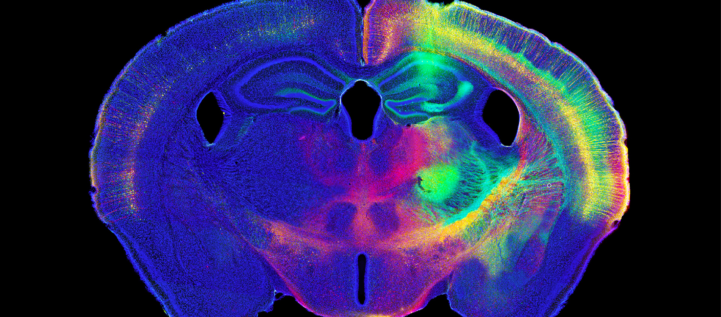
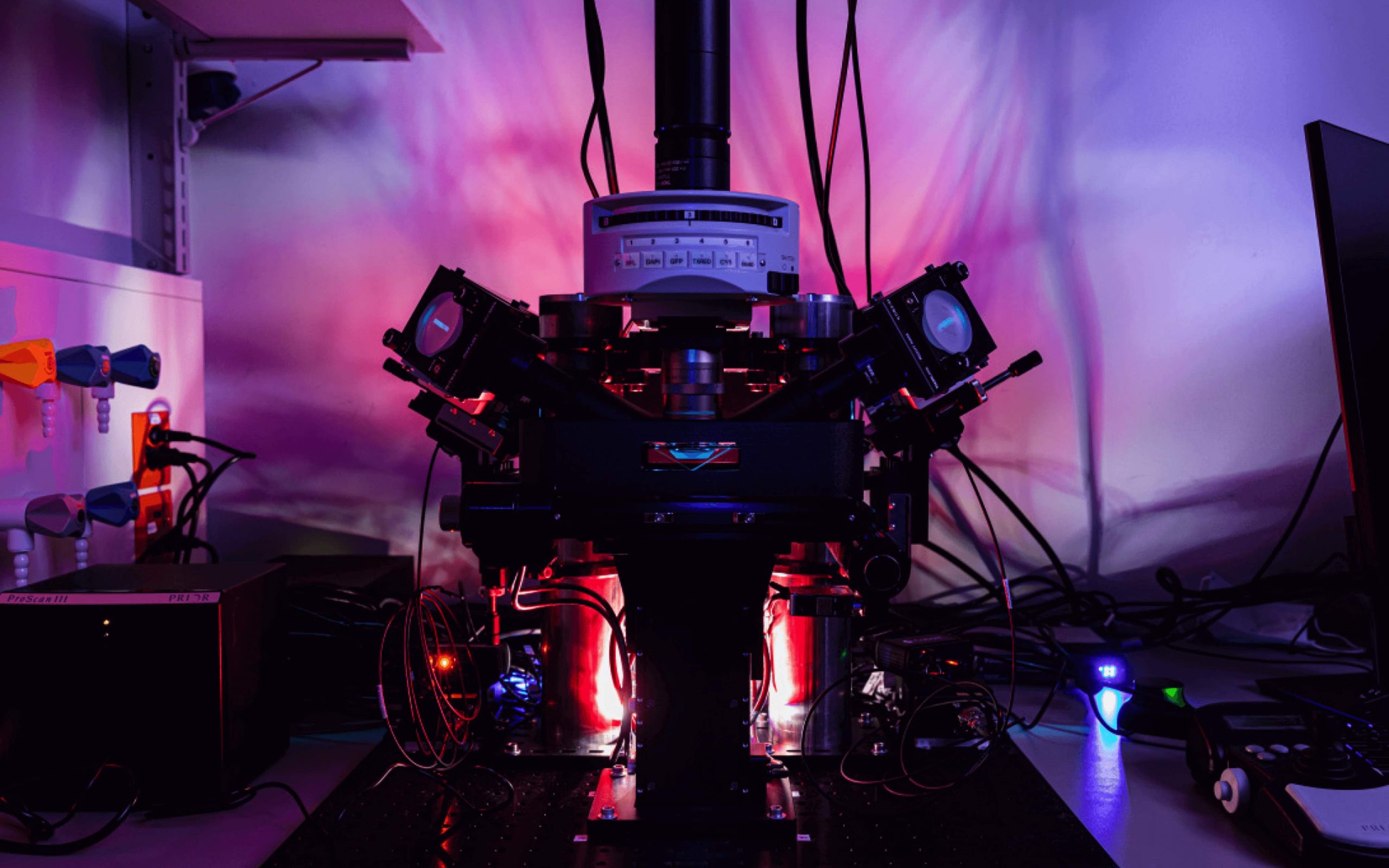
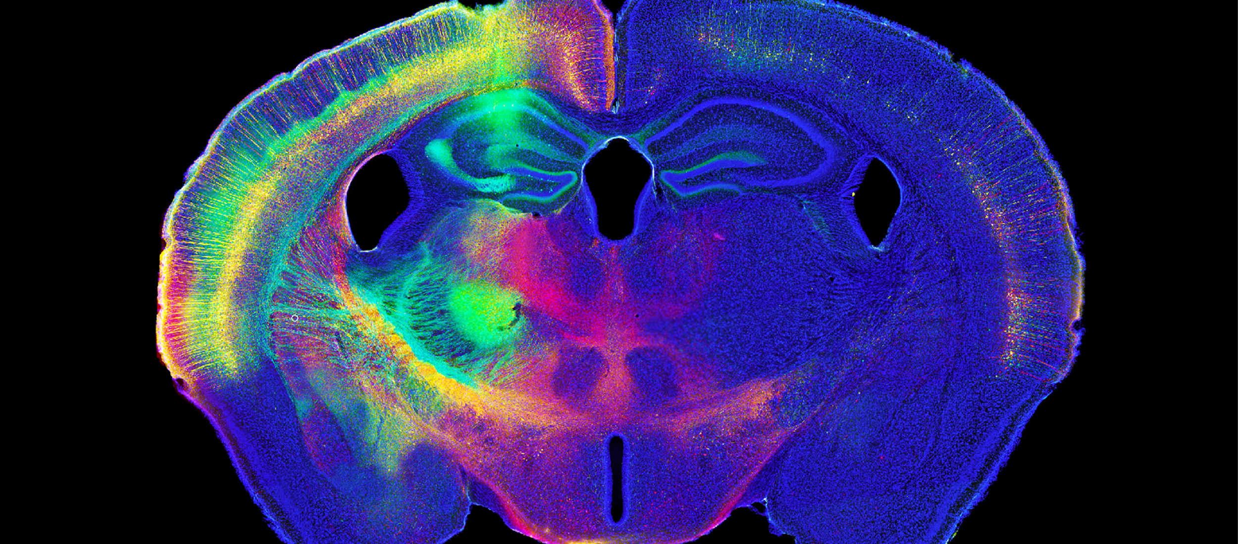
For Immediate Release: Williston, VT (September 25, 2020)—To support the scientific community’s movement toward a more open and collaborative climate, MBF Bioscience is making their digital reconstruction file format, the Neuromorpholocial File Specification open and available to all researchers. MBF Bioscience has a long history of supporting openness and collaboration in neuroscience research. MBF Bioscience’s file format has evolved for over 30 years, influenced by leading neuroscientists,...
Read More
MBF Bioscience Blog
Scientists at Western Sydney University used Stereo Investigator and Neurolucida 360 to quantify cells in a mouse model of neuroinflammation after feeding mice two different curcumin formulations. Some inflammation is normal in a healthy mammalian brain. But as the brain ages, processes can break down, leading to chronic neuroinflammation. This can develop into Alzheimer’s disease, dementia, and other neurodegenerative diseases. Scientists at Prof. Gerald Muench’s lab, at...
Read MoreMBF Bioscience was just awarded a patent by the USPTO for our new brain mapping technology! This novel technology is now available in our BrainMaker and NeuroInfo software to map image data and all associated measurements into a common coordinate system so that data can be compared across animals, cohorts, and laboratories. Learn more about NeuroInfo at https://www.mbfbioscience.com/neuroinfo ...
Read MoreResearchers Quantify Improvement in Heart Vasculature with Vesselucida 360 and Vesselucida Explorer Cells need oxygen to survive, but during a heart attack, blood flow is restricted and cardiac cells can’t get the oxygen they need to stay alive. A new therapy, developed by researchers at the Coulombe Lab at Brown University may be able to provide the heart with the support it needs to recover after...
Read MoreBiolucida for Medical Education is a learning platform that serves high resolution microscope slides to students without the need for a physical microscope. As universities around the world move to online classes during the COVID-19 pandemic, educators suddenly need to find ways to create authentic learning experiences for their students. Biolucida for Medical Education creates that experience by giving students studying at home the same experience...
Read MoreAn interdisciplinary team of researchers, including MBF Bioscience’s Dr. Susan Tappan and Maci Heal, have created a fully reconstructed, virtual 3D heart, digitally showcasing the heart's unique network of neurons for the first time. The investigators in this study--appearing May 26 in the journal iScience--created a comprehensive map of the intrinsic cardiac nervous system at a cellular scale using MBF Bioscience’s Tissue Mapper and TissueMaker software....
Read MoreNeurolucida 360 Used to Analyze Dendrites and Dendritic Spines Amyloid plaques and tau tangles are the hallmarks of Alzheimer’s disease (AD) pathology, but synapse loss is what causes cognitive decline, scientists say. In a paper published in Science Signaling, researchers at the Herskowitz Lab, at the University of Alabama at Birmingham, used Neurolucida 360 to analyze spine density and dendritic length in hAPP mice — a...
Read MoreFor Immediate Release: Williston, VT (February 04, 2020) — MBF Bioscience’s revolutionary light sheet microscope system, ClearScope, sets a new standard for microscopic imaging. The new decade is poised to bring about incredible scientific innovations, and MBF Bioscience is leading the charge in 2020 with the creation of the “light sheet theta microscope” system, ClearScope. MBF Bioscience secured exclusive license from Columbia University to develop the light...
Read MoreNew Software Application Quantifies Changes in Dendritic Spine Morphology Over Time Williston, VT — December 10, 2019 — The ability to track the changes that occur in dendritic spine morphology over time is critical to many scientific studies, which is why MBF Bioscience is pleased to announce the launch of MicroDynamix. This powerful new software application helps neuroscientists acquire more information about morphological changes in the...
Read MoreScientists use NeuroInfo to help navigate the brain and compare findings across labs Reproducibility has always been a primary goal in science. But the human effort involved in replicating a research study and analyzing the results, can be considerable. NeuroInfo®is a revolutionary new tool that scientists are using to register whole slide images into a standardized mouse brain atlas in an easy, automated way. Images...
Read More


