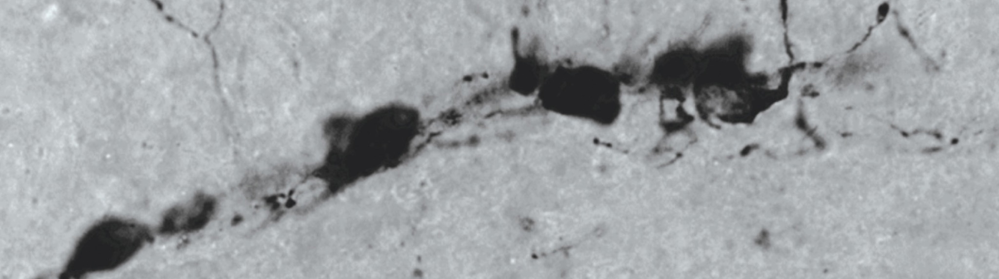
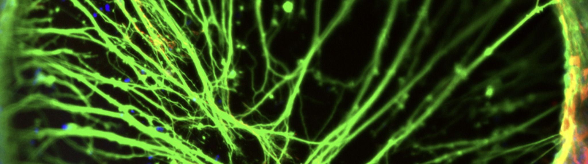
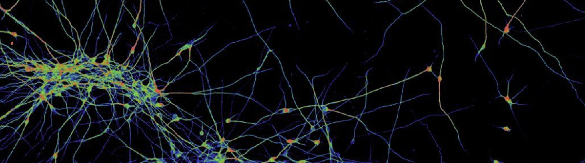
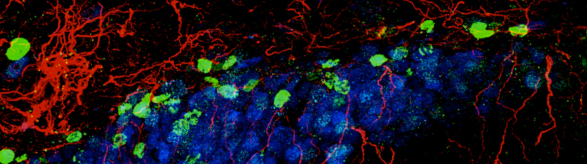
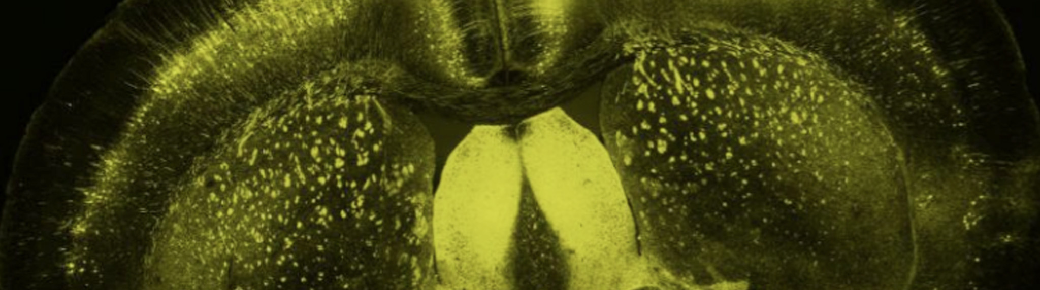
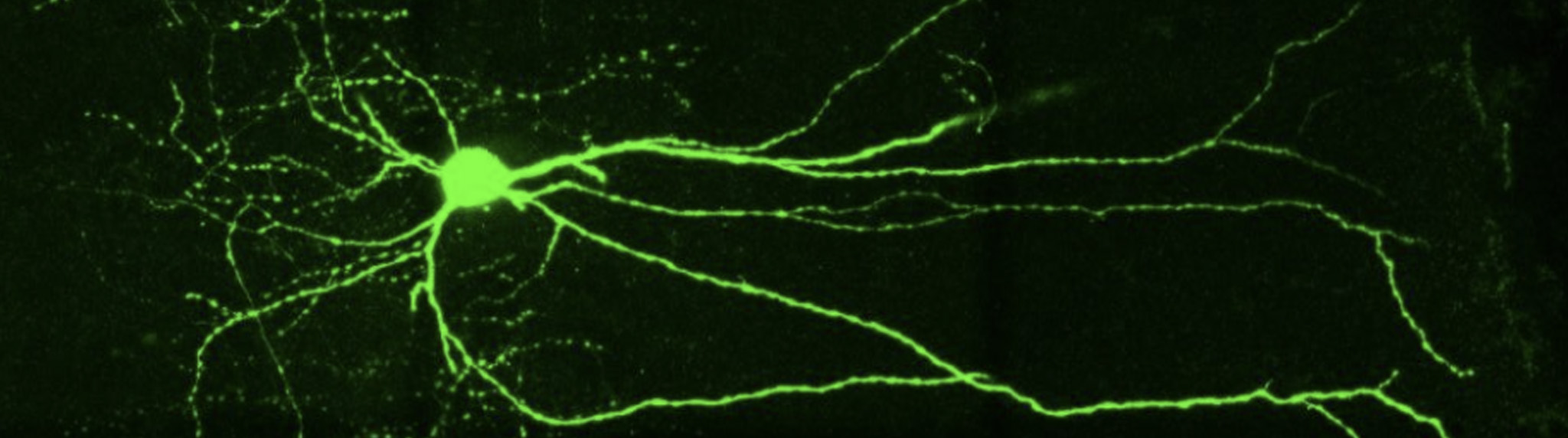
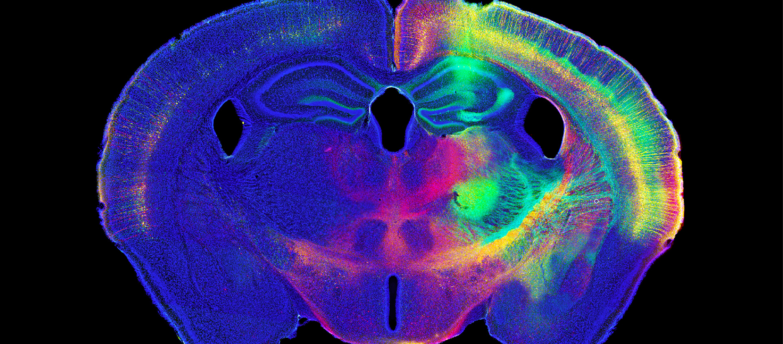
MBF Bioscience Blog

  According to scientists at the Hotchkiss Brain Institute in Calgary, Canada, there is evidence for increased neurogenesis in adult mice reared by two parents. Their study also describes other interesting findings, such as the fact that increased neurogenesis persists in the next generation, or that the effects of differences in rearing affect males and females differently.   Using Stereo Investigator to perform a stereological assessment of proliferating cells...

Read More

Researchers at the Waisman Center (University of Wisconsin-Madison) just took a big step in their quest to develop regenerative medicines for treating Parkinson's, Alzheimer’s, and other neurodegenerative diseases. They used human embryonic stem cells to restore memory and learning in disabled mice.   The study, published last month in Nature Biotechnology, "is the first to show that human stem cells can successfully implant themselves in the brain...

Read More

MBF Bioscience received a prestigious Tibbetts Award during a ceremony at the White House on May 16. The Tibbetts Award recognizes MBF Bioscience for a number of accomplishments including: technological innovation, serving federal Research and Development needs, encouraging diverse participation, and increasing the practical commercial use of federal research.   The Tibbetts Award is given to companies who participate in the Small Business Innovation Research program which is run by...

Read More

Spinal cord injuries can result in a range of physical disabilities from slight loss of motor function to major paralysis, but little is known about the mechanisms underlying the damage. Scientists affiliated with the Miami Project to Cure Paralysis at the University of Miami are gaining knowledge about how the nervous system responds to spinal cord injuries. Their latest study, published last month in the...

Read More

  What does it take to survive that tumultuous time called adolescence? Good friends, exercise, and new brain cells.   Scientists at Michigan State University found evidence of neurogenesis in the brains of adolescent hamsters, according to a study published last month in PNAS. The new cells, which became integrated into neural circuits in adulthood, were discovered in the amygdala and connected limbic regions – areas associated with...

Read More

  The importance of studying the brain in three dimentions is something we understand at MBF Bioscience. Every day scientists around the world use our products to reconstruct neurons and analyze brain cells in 3D. That's why we're excited to hear about the new possibilities for whole brain analysis coming out of Dr. Karl Deisseroth's lab at Stanford University.   A press release issued last week describes a...

Read More

There's a lot to be said for being in the right place at the right time. For a neuron, emerging at a certain place within the brain destines it for a particular function. A new study posits that, for a group of cells in the hippocampus, it's not only where a neuron is born, but also when it is born, that defines the specific roles...

Read More

Microscopes and glass slides are on their way to becoming obsolete in medical and science courses. MBF Bioscience just unveiled Biolucida Cloud 2.0, software that simulates the experience of using a light microscope, complete with the ability to focus through specimens. This software enables educators to integrate virtual slides — high-resolution digital images of specimens mounted on glass slides — into their course materials and tests.   Thanks to...

Read More

A stroke patient is rushed to the hospital. Deprived of oxygen-rich blood, brain cells have already died, and more damage will probably occur in the hours and days to come. But researchers at the University of South Florida and the University of Padova in Italy say a two-part package administered through the body, rather than directly into the brain, may be the key to staving...

Read More

[caption id="" align="alignright" width="189"] Overall view of the computer microscope developed by Drs. van der Loos and Glaser (circa 1965).[/caption] Prior to the computer microscope era, quantitative neuro-anatomical studies were performed using the camera lucida method, an optical method allowing the scientist to see the neurons as if reflected on the piece of paper on which she will trace. These studies were painstaking and extremely time-consuming....

Read More