
Exercise changes astrocytes and eases symptoms of neurodegenerative disorders
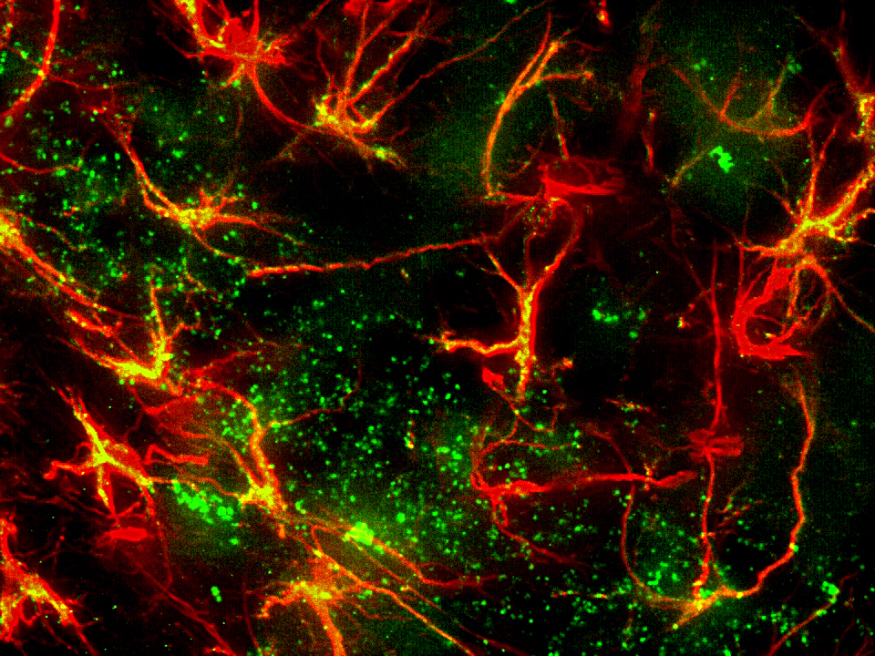
Astrocytes (GFAP) in the dentate gyrus of a mouse hippocampus. Image courtesy of Dr. Ahmad Salehi, Stanford University.
It is well known that physical exercise eases the symptoms of neurodegenerative disorders like Alzheimer’s disease and helps to prevent their onset. Researchers at Stanford University are working on figuring out how it happens.
In their study, published in the journal Brain Structure and Function, scientists in Dr. Ahmad Salehi’s lab examined the effects of physical exercise on astrocytes in a region of the mouse brain that is critical for cognition – the dentate gyrus of the hippocampus. Previous studies have shown that an increase in the expression of brain-derived neurotrophic factor (Bdnf) occurs in this region after exercise (Philips, Salehi et al 2014). Bdnf is a protein that supports the survival of existing neurons and encourages new growth, playing an important role in cognitive function.
While the current study reconfirms that exercise generates increased levels of Bdnf (more than a fourfold increase in exercised mice versus non-exercised mice), it also describes several new findings including increased synaptic load in the dentate gyrus, alterations in the morphology of astrocytes, and changes in the orientation of astrocytic projections toward dentate granule cells.
The authors speculate that the changes they observed may be attributed to increased expression of a receptor called TrkB, which astrocytes express in response to increases in Bdnf levels. According to the paper, TrkB binds to Bdnf, activating the mechanisms behind neuronal development.
“Our study suggests that astrocytes actively respond and could indeed mediate the positive effects of physical exercise on the central nervous system and potentially counter degenerative processes during aging and neurodegenerative disorders,” (Fahimi, et al 2016).
The researchers used Neurolucida to determine the location, the extent, and orientation of astrocytic projections, finding a significant increase in the length of astrocytic projections in exercised mice.
“Neurolucida is one of the very few systems that combines complex morphometrical quantification with beautiful display of the results,” said Dr. Salehi, Clinical Professor, Department of Psychiatry and Behavioral Sciences at Stanford Medical School.
Since astrocytes help prevent excitotoxicity in the brain by removing excess glutamate from extracellular space, the researchers speculate that the increased length of astrocytic projections they observed in exercised mice could make this process more efficient.
Differences in the orientation of astrocytic projections were also reported, with the majority of projections of exercised mice directed toward the dentate granule cell layer – a region featuring increased levels of Bdnf release and synthesis after exercise.
The number of astrocytes in the molecular layer of the dentate gyrus in exercised and non-exercised mice was quantified with Stereo Investigator, however, there was no significant difference in astrocyte populations between the two groups.
“In summary, our study suggests that astrocytes constitute an important element in mediating the positive effects of physical exercise in the dentate gyrus of the hippocampus. Furthermore, it appears that physical exercise-induced release of Bdnf by the DG leads to a significant alteration in structure and function of astrocytes in protection against glutamate toxicity during aging and a number of neurodegenerative disorders,” (Fahimi et al 2016)
Fahimi, A., Baktir, M.A., Moghadam, S., Mojabi, F.S., Sumanth, K., McNerney, M.W., Ponnusamy, R., Salehi, A. Brain Struct Funct (2016). doi:10.1007/s00429-016-1308-8
Phillips, C., Baktir, M.A., Srivatsam, M., Salehi, A. Front. Cell. Neurosci., (2014) https://doi.org/10.3389/fncel.2014.00170