Scientists Observe Differences Between Brains of Stressed and Unstressed Rats After Fear Conditioning
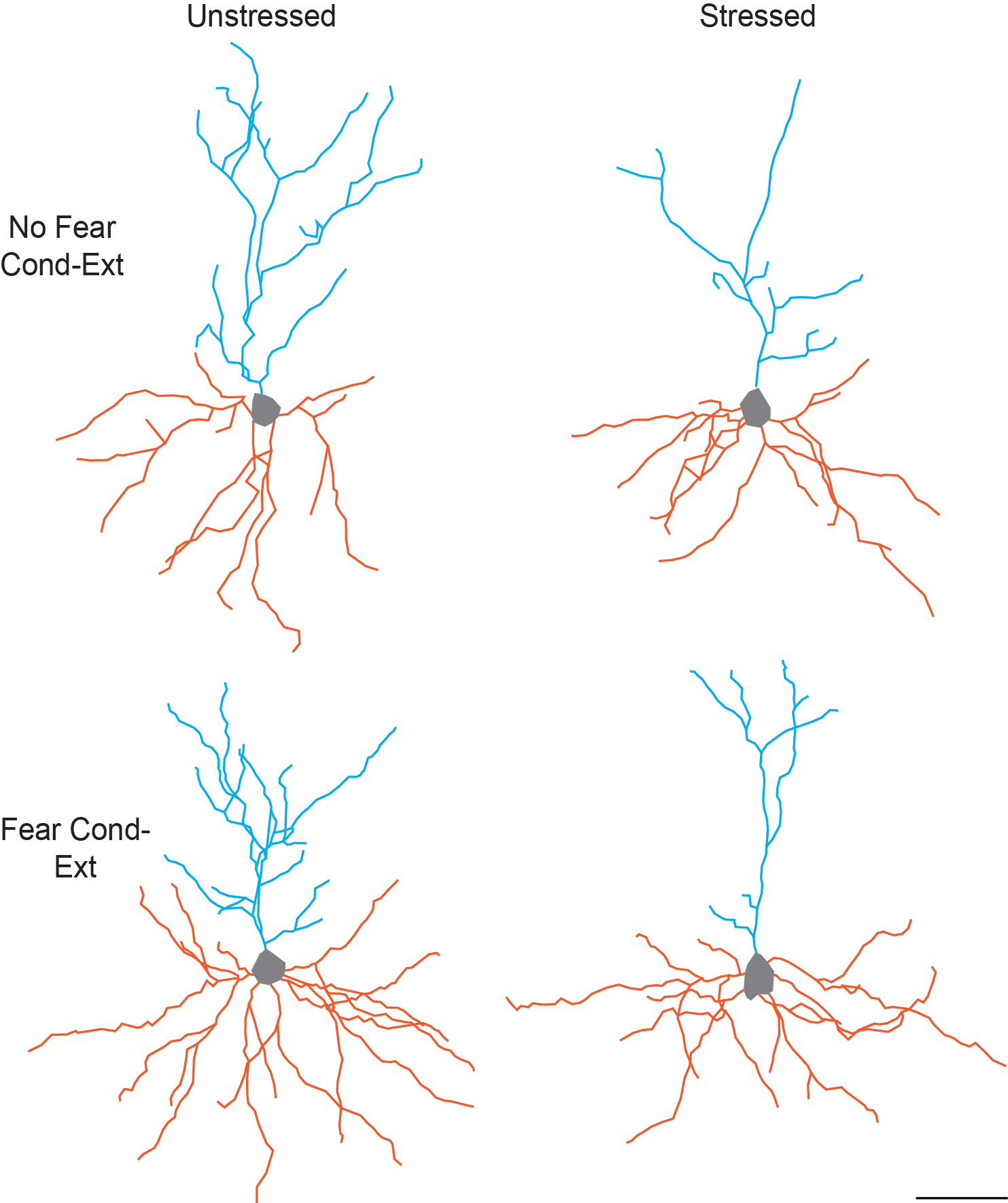
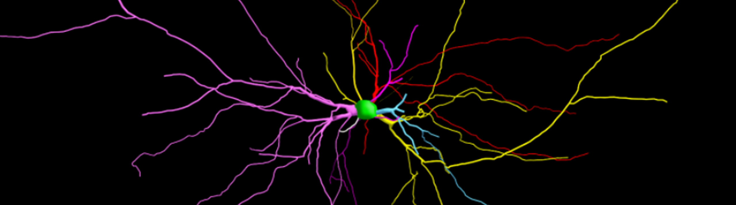
This figure illustrates the separate and combined effects of acute stress and fear conditioning/extinction on dendritic morphology of pyramidal neurons in the infralimbic region of medial prefrontal cortex. Each neuron shown is a composite made up of apical (blue) and basilar (orange) arbor near the mean of the group. The apical and basilar arbors of each composite are from different neurons. Image courtesy of Cara Wellman, PhD.
A soldier jumps at the sound of fireworks. Though there is no threat to his or her life, the blasts mimic the ones heard on the battlefield, and that fear response is not easy to forget. The process of shedding a fear response like this one is called fear extinction. Scientists think patients suffering from stress-sensitive psychopathologies, like Post-Traumatic Stress Disorder, aren’t able to suppress certain fear responses because of deficits in their brain circuitry induced by stress.
A recent study by researchers at Indiana University and the University of Haifa, in Israel, describes significant differences between the brains of stressed rats and unstressed rats.
Using Neurolucida to analyze neurons in the infralimbic cortex (IL) – a region of the brain associated with fear extinction – the research team found that stressed rats had shorter dendrites and less dendritic branching in pyramidal neurons of the IL. They also found that while stress had no affect on spine density, rats that underwent fear conditioning and extinction had decreased spine density on apical terminal branches, providing evidence that dendritic morphology in this region is sensitive to stress, while spine density may be a reflection of learning.
“Having helped colleagues set up procedures for neuron reconstructions and spine counts in labs that aren’t equipped with Neurolucida, I can tell you with complete confidence that my lab wouldn’t be nearly as productive without our Neurolucida system,” said Dr. Cara Wellman. “It makes mapping out regions of interest, identifying neurons for reconstruction, and reconstructions, and data analysis a simple and streamlined process. My students and I especially appreciate the Lucivid, which allows us to trace neurons while looking through the oculars – so much easier and clearer in my opinion than on a video monitor.”
To achieve their results, the researchers subjected rats to fear conditioning, where they learned to associate a certain tone with a footshock. Some of the rats were then exposed to an elevated platform in a brightly lit room for 30 minutes (stressed) while others returned to their home cages (unstressed). Next came extinction sessions. In a test to see if they would be able to shed the fear response associated with the stimulus, rats were placed in a space where they heard a tone but did not experience a footshock. The scientists observed that stressed rats exhibited freezing during the extinction sessions at a much higher rate than unstressed rats, leading them to believe that rats exposed to acute stress were resistant to fear extinction.
Further quantification of apical and basilar dendritic branching in the pyramidal neurons of the IL, measured with three-dimensional Sholl analysis, confirmed differences between the stressed and unstressed rats’ brains that correlated with fear behavior.
“The main findings of the current study were that acute stress, concurrent with producing resistance to extinction, produced changes in morphology of pyramidal neurons in IL,” the authors say in their paper. “These findings provide evidence that alterations in IL pyramidal neuron morphology occur quickly and differentially in response to acute stress and fear conditioning/extinction.”
Moench KM, Maroun M, Kavushansky A, Wellman C. Alterations in neuronal morphology in infralimbic cortex predict resistance to fear extinction following acute stress. Neurobiology of Stress. 3: 23-33. doi:10.1016/j.ynstr.2015.12.002