
Stereological Study Reveals Neuron and Glia Proliferation in Hippocampus of Lithium-Treated Mice
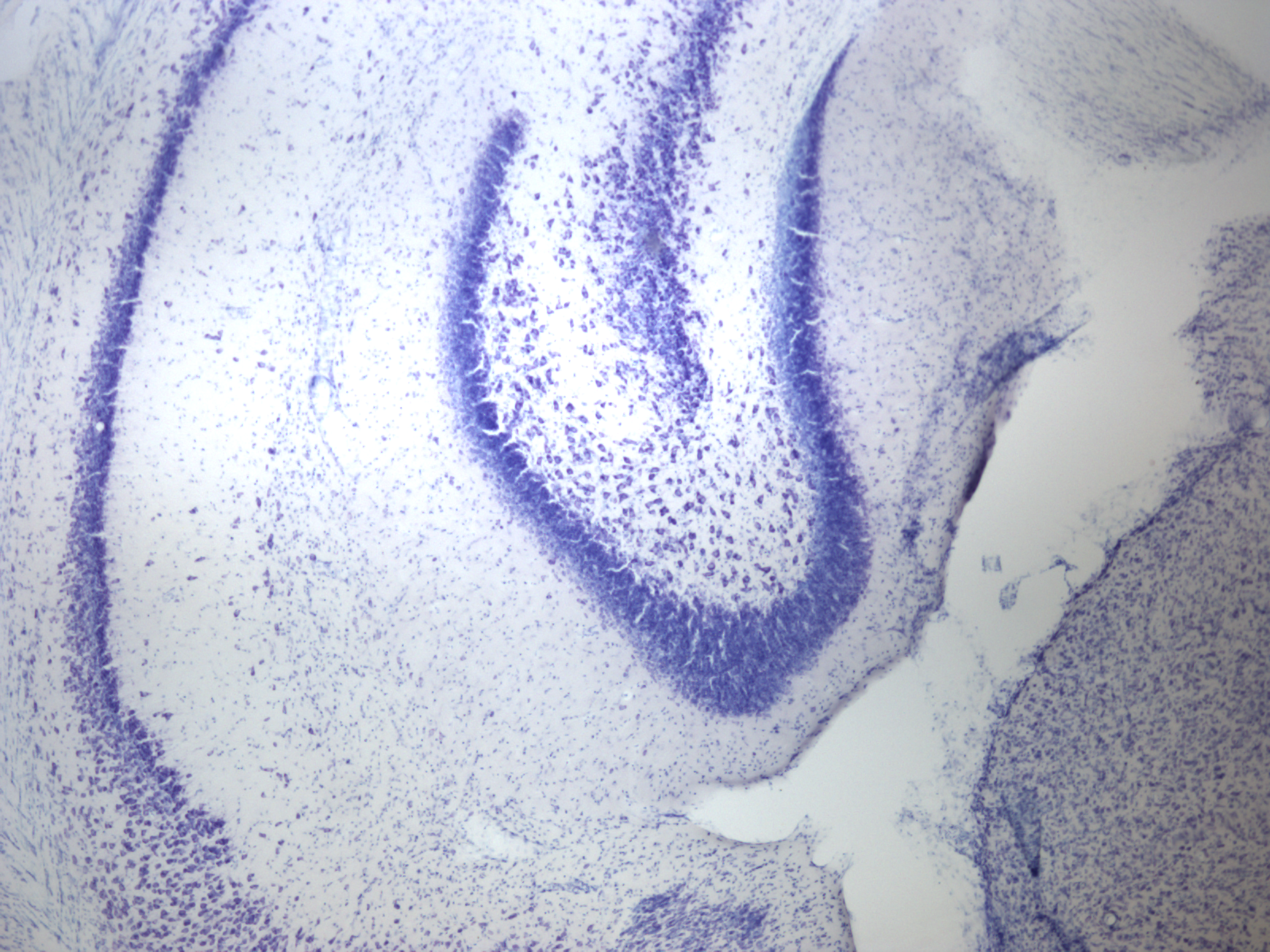
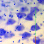
The optical fractionator probe was used to quantify the number of neurons and glia in the dentate gyrus
Doctors have used lithium to treat patients with bipolar disorder since the 1970s. Known for its efficacy in stabilizing patients’ moods by regulating manic episodes, lithium is also associated with a decreased risk of suicide. But while this naturally occurring element is the most widely prescribed medication for those suffering from bipolar disorder, scientists still have much to learn about how lithium physically affects the brain.
A recent study published in the journal Bipolar Disorders adds to the growing body of evidence that says lithium contributes to cell proliferation in parts of the brain. Conducted by scientists at the University of Mississippi and the VU University Medical Center in Amsterdam, the study revealed an increased number of neurons and glia, and increased astrocyte density in the dentate gyrus of lithium-treated mice versus controls treated with a placebo.
Using the optical fractionator probe in Stereo Investigator, the researchers quantified the number of Nissl stained neurons and glial cells, and calculated astrocyte density. The results showed twenty-five percent more neurons and twenty-one percent more glia in the denate gyrus of lithium-treated mice. They also performed a stereological examination of another brain region – the medial prefrontal cortex (mPFC), but did not witness significant differences between lithium-treated and control mice in this area.
“In this study, particular cortical regions, ie. the fascia dentata in the hippocampus and the mPFC in the cerebral cortex needed to be selected in histological sections of the mice brains,” explained Dr. Harry B.M. Uylings, “therefore the stereological counting procedure applied was the best one. Stereo Investigator greatly assisted in the counting of cells, and the software’s excel data-output was especially beneficial.”
According to the paper, the findings present a more detailed picture of lithium-induced alterations in the dentate gyrus cellular phenotype than previously available, and provide the first evidence for lithium-induced increases in glia and astrocytes.
The authors also explain that while cell number increased in the dentate gyrus of lithium-treated mice, the region’s overall volume as well as that of the greater hippocampus was unaffected by the element. The volume of the dentate gyrus and the hippocampus as a whole was measured with the Cavalieri method in Stereo Investigator. The researchers describe the dissociation between cell proliferation and volume as “an interesting observation that warrants further investigation.”
Rajkowska, G., Clarke, G., Mahajan, G., Licht, C.M., van de Werd, H.J., Yuan, P., Stockmeier, C.A., Maji, H.K., Uylings, H.B., Differential effect of lithium on cell number in the hippocampus and prefrontal cortex in adult mice: a stereological study. Bipolar Disord. 2016 Feb;18(1):41-51. doi: 10.1111/bdi.12364.