Software Applications For Quantitive Analysis

MBF Bioscience >  Blog > MBF Products & Service Solutions  > Software Applications For Quantitive Analysis (Page 3)

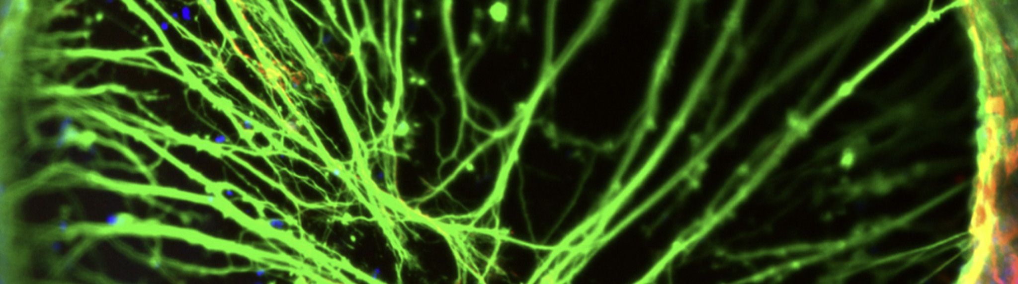
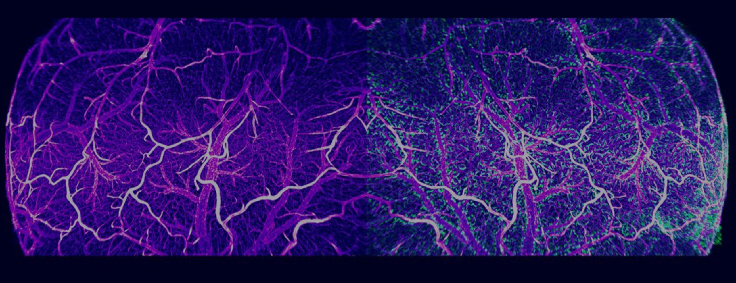
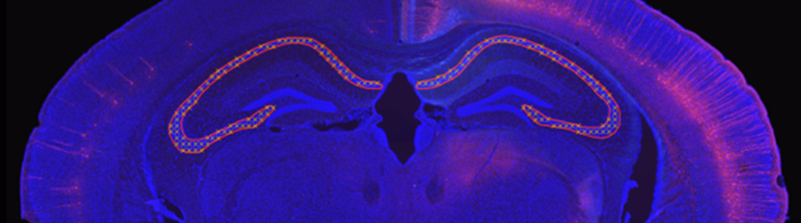
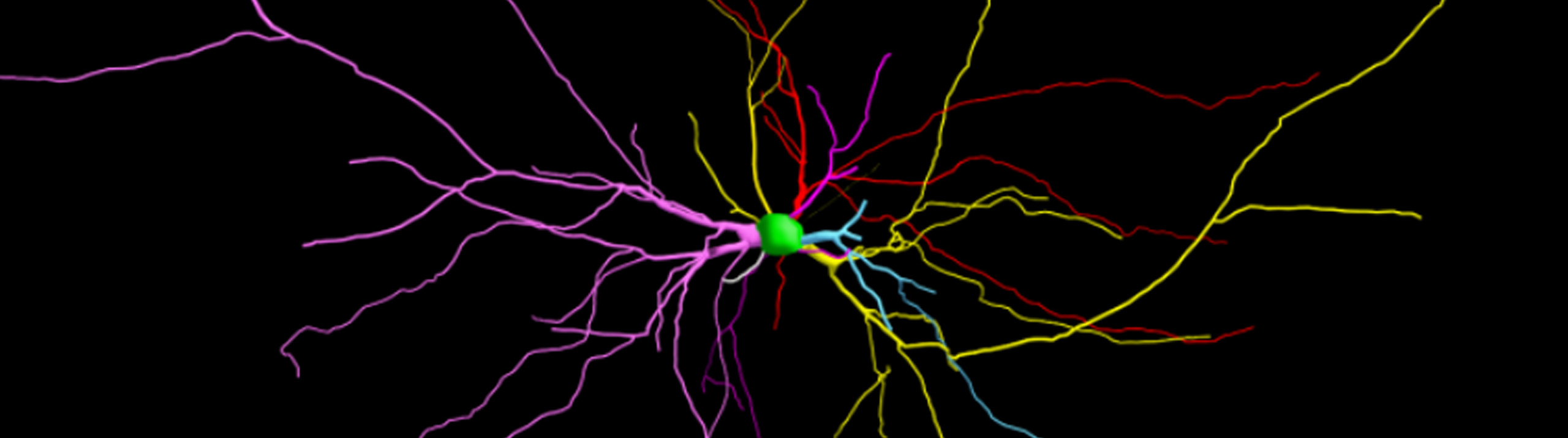
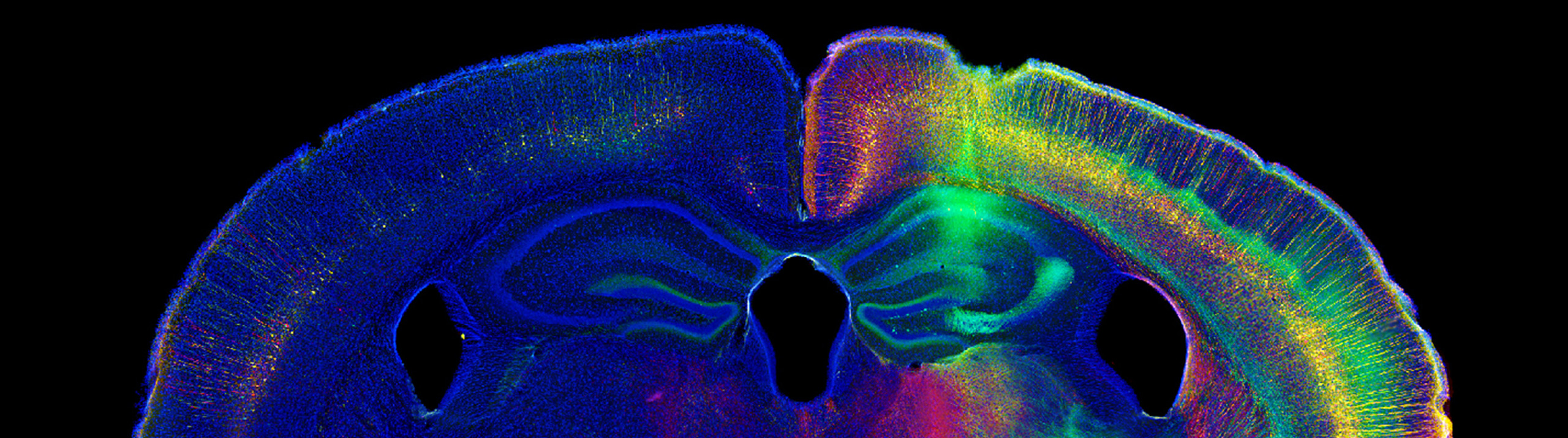
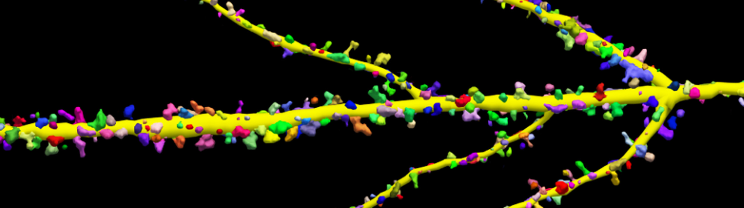
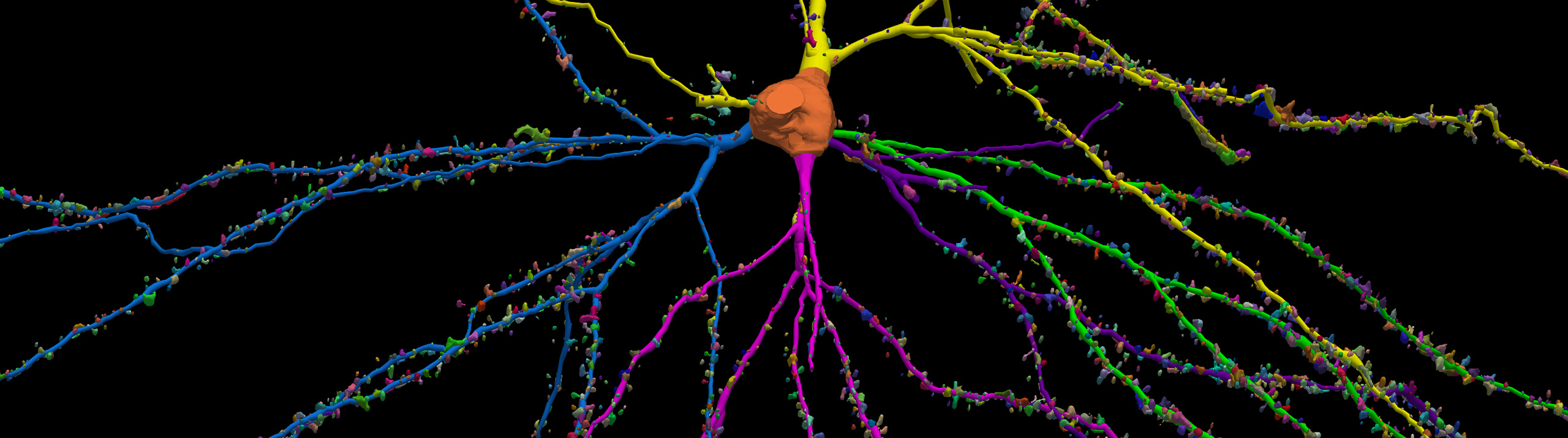
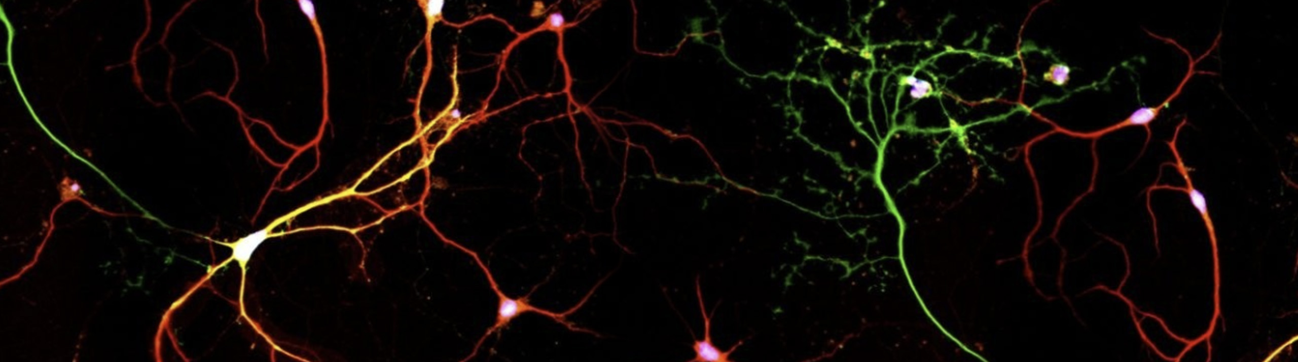
Combination of new microscopy and expansion tissue preparation methods facilitate better and faster analysis of subcellular neural elements. Today, the journal Science published a paper authored by a research team led by Dr. Ed Boyden of MIT and Nobel Prize recipient Dr. Eric Betzig of Janelia Research Campus. Among the authors are MBF Bioscience Scientific Director Dr. Susan Tappan and Senior Software Engineer Alfredo Rodriguez. In the...

Read More

FOR IMMEDIATE RELEASE MBF Bioscience Williston, VT – January 9, 2019 – MBF Bioscience is pleased to announce our participation in the Stimulating Peripheral Activity to Relieve Conditions (SPARC) program. Funded by the National Institutes of Health (NIH), this extensive research initiative is a vast collaborative effort, which aims to deepen the understanding of how the peripheral nervous system impacts internal organ function.   “We are honored to be working...

Read More

MBF Bioscience now offers customized models of Huron Digital Pathology’s TissueScope, a line of whole slide scanners, and supports TissueScope images across its range of analysis software. October 23, 2018 – Huron Digital Pathology and MBF Bioscience are proud to announce their partnership to offer customized models of Huron’s TissueScope whole slide scanners integrated with MBF’s Stereo Investigator®- Whole Slide Edition, NeuroInfo®, Biolucida®, and BrainMaker®...

Read More

Neurolucida and Neurolucida Explorer Used for 3D Reconstruction and Quantitative Analysis Researchers used Neurolucida to reconstruct a newly discovered type of neuron found only in the human brain, according to a study published in the journal Nature Neuroscience. Known as “rosehip” neurons because of the way they resemble a rose after its petals have fallen off, these cells feature compact, bushy axonal arborizations.   Found in the first...

Read More

Williston, VT – September 5, 2018 – Researchers studying microvascular networks and vessels have a groundbreaking new software application to facilitate their work. Developed by MBF Bioscience, Vesselucida®360 automatically reconstructs and analyzes microvascular networks in 3D.   Specifically designed to recognize the intricacies of the vascular system, Vesselucida features sophisticated algorithms that quickly and accurately create 3D reconstructions of images and tissue specimens. Built-in analyses provide data...

Read More

Williston, VT – June 29, 2018 – MBF Bioscience is pleased to announce the launch of the Whole Slide Edition of Stereo Investigator. This is a new version of our renown Stereo Investigator software, designed especially for conducting stereology on images from slide scanners.   Featuring a streamlined, user-friendly interface, this new product includes a variety of probes for quantifying number, length, volume, and surface area of...

Read More

[caption id="attachment_6839" align="aligncenter" width="632"] Image Courtesy: Bob Jacobs, Ph.D. , Colorado College[/caption]   With the release of its new version on November 28, NeuroMorpho.org adds 9,987 new images to its archive, bringing its impressive collection of digitally reconstructed neurons to 80,012.   Scientists used MBF Bioscience’s software, Neurolucida and Neurolucida 360, to reconstruct the majority of these cells. In fact, 64 times more neurons were reconstructed with MBF Bioscience...

Read More

[caption id="attachment_6830" align="aligncenter" width="632"] An experimental coronal mouse brain section automatically aligned to the Allen Mouse Brain Reference Atlas[/caption]   Analyzing cellular populations within specific anatomies in brain images requires expertise in both neuroanatomy and cellular identification. This typically involves a scientist comparing experimental images with a reference atlas and manually delineating anatomical regions and marking cell populations within. NeuroInfo®, a revolutionary new technology from MBF Bioscience,...

Read More

Following a well-designed protocol is essential to achieving accurate and consistent results in scientific research. Now, scientists using Neurolucida 360 for dendritic spine and neuron analysis can follow a published set of guidelines to ensure optimal confocal data series for proper dendritic spine quantification and neuron reconstruction. The paper, written by MBF Bioscience scientists and researchers from the Icahn School of Medicine at Mount Sinai...

Read More

The 3 algorithms in Neurolucida 360 were used in combination to create a smooth, accurate reconstruction   Minor releases typically don't include new features, but Neurolucida 360 isn't an ordinary piece of software. Neurolucida 360 v2.7 has many new features and improvements, including: A new automatic tracing algorithm - Rayburst Crawl Capture videos of your rotating neuron reconstructions for presentations and publications New backbone length analysis for dendritic spines Improved handling of images...

Read More