Researchers quantify cortical cell numbers in cleared tissue with new unbiased stereology technique
The Image Volume Fractionator probe, available in Stereo Investigator – Cleared Tissue Edition, is facilitating huge efficiency gains for quantifying the number of cells.
At Dr. Patrick R. Hof’s lab at the Icahn School of Medicine at Mount Sinai, researchers imaged the cerebral cortex using light-sheet fluorescence microscopy and quantified the number of neurons, including those that express proteins involved in Alzheimer’s disease and schizophrenia, using the Image Volume Fractionator1. This work marks the beginning of an important and ambitious project to build an atlas of cortical cells, using a multi-resolution imaging pipeline. At the pipeline’s highest level of resolution, both the Image Volume Fractionator, for use with thick sections of cleared tissue, and the Optical Fractionator, for much thinner sections, are being used to estimate cell number. The researchers will also use automatic cell detection and plan to compare results obtained using the three methods.
In the paper A Multimodal Imaging and Analysis Pipeline for Creating a Cellular Census of the Human Cerebral Cortex1, the authors describe the beginning of the effort to build a census of the human cerebral cortex, a laminar structure, that contains layers comprised of different cell types visible using high resolution microscopy. There are a number of different neuronal cell types in each layer, including projection neurons and interneurons, as well as excitatory and inhibitory neurons. Layers can be identified based on different proteins contained in certain cells using fluorescence immunohistochemistry. Calretinin, a calcium-binding protein, is found in a subpopulation of the inhibitory interneurons that contain GABA. Neurofilament protein, which can be found in the cytoskeleton, makes up 30 percent of cortex cells. Parvalbumin, another calcium-binding protein, is also found in a subset of cortical cells.
The cells in the cortex have a purpose that is supported by their neurochemical and anatomical characteristics. Here are two examples involving Alzheimer’s disease and schizophrenia. Calretinin-positive cells in the cortex are spared in Alzheimer’s disease2, but neurofilament protein-positive cells degenerate, and that degeneration may predict cognitive decline3. The parvalbumin-containing basket cell is an inhibitory GABAergic interneuron in the cortex that inhibits the main output cell—the pyramidal neuron. Problems with this cell type may affect gamma oscillations, leading to the deficits in cognitive control that accompany schizophrenia4.
Wouldn’t it be valuable to have an atlas or census of the cortex that is “zoomable” like a GPS map, and shows the cell types and their connections? Hof. et.al., demonstrate that this is possible using three imaging techniques at increasing resolutions (Fig. 1).
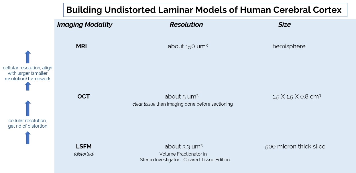
Fig. 1 The three imaging modalities used in this study. Magnetic Resonance Imaging (MRI) is the lowest resolution. Optical Coherence Tomography (OCT) is the mid-resolution. Light Sheet Fluorescence Microscopy (LSFM) is the highest resolution. The Image Volume Fractionator probe is carried out using LSFM. Those images are registered to eliminate distortion to help match them to OCT and the MRI images.
At the most highly resolved level, LSFM, two unbiased stereology techniques are used to build a census inside the atlas: the Optical Fractionator and the new, Image Volume Fractionator. The latter is made possible by tissue clearing methods, which in turn allows for the use of tissue sections that are, in this case, 10 times thicker than for the Optical Fractionator (Fig. 2).
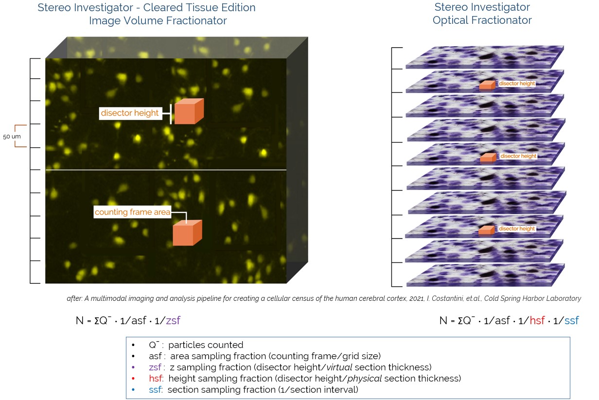
Fig. 2 The Image Volume Fractionator (IVF) was designed to be used on thick sections or large intact specimens. It is much more efficient than working on traditional histological sections that were not cleared and therefore need to be, in this case, 10 times thinner. N is the estimate of number of cells. Systematic random sampling and disector rules are followed while counting.
Since the LSFM images are ten times thicker than the thinner sections needed in the absence of tissue clearing, counting with the Image Volume Fractionator probe can be done more quickly. It is much more efficient to count cells in one large image than in ten separate thinner tissue sections. There is also less sectioning artifact, which helps with registering the higher resolution LSFM images back to the larger volume MRI images.
We are excited to see this new use of the Image Volume Fractionator, which increases efficiency and reduces imaging distortions from physical sectioning. The potential that cleared tissue offers for increasing efficiency is great, but is still largely untapped. As this method is used more frequently, we look forward to hearing feedback from the research community to further improve the capabilities and usability of the Image Volume Fractionator in Stereo Investigator – Cleared Tissue Edition.
References:
1) A Multimodal Imaging and Analysis Pipeline for Creating a Cellular Census of the Human Cerebral Cortex 2021, Constantini, et al., https://www.biorxiv.org/content/10.1101/2021.10.20.464979v1
2) Hof, P. R., Nimchinsky, E. A., Celio, M. R., Bouras, C. & Morrison, J. H. Calretinin, Immunoreactive neocortical interneurons are unaffected in Alzheimer’s disease. 861 Neurosci Lett 152, 145-148 (1993).
3) Bussiere, T. et al. Progressive degeneration of nonphosphorylated neurofilament protei enriched pyramidal neurons predicts cognitive impairment in Alzheimer’s disease: Stereologic analysis of prefrontal cortex area 9. Journal of Comparative Neurology (2003).
4) Glausier, J. R., Fish, K. N. & Lewis, D. A. Altered parvalbumin basket cell inputs in the dorsolateral prefrontal cortex of schizophrenia subjects. Mol Psychiatry 19, 30-36 (2014). Lewis, D. A., Curley, A. A., Glausier, J. R. & Volk, D. W. Cortical parvalbumin interneurons and cognitive dysfunction in schizophrenia. Trends Neurosci 35, 57-67 (2012).