
Researchers Map and Explore the Heart’s “Little Brain”
The heart has a “little brain.” It’s a network of neurons known as the intrinsic cardiac nervous system (ICNS), and it plays a key role in regulating cardiac activity.
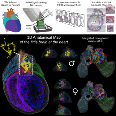
Building on previous research (Achanta et al., 2020), which resulted in a 3D map of the rat ICNS, a new study by a team of scientists from the University of Central Florida, Thomas Jefferson University, the University of Auckland, Strateos Inc, and MBF Bioscience further explores the organization of peripheral neurons in the ICNS of the rat heart and describes variations in female and male rats.
Using MBF Bioscience’s Tissue Mapper® software, the research team, which included Scientific Director Dr. Susan Tappan, and Research Analyst Maci Heal, comprehensively annotated, mapped, and visualized the anatomy and ICNS of female and male rat hearts in 3D.
By marking the ICNS neuron locations for each heart using Tissue Mapper, the investigators found that the pattern, distribution, and clustering of ICNS neurons across all male and female rat hearts is highly conserved and neuron clusters are organized and localized. Female hearts had fewer neurons, lower packing density, and slightly reduced distribution than male hearts.
“The team at MBF has been highly receptive to our research needs,” says Dr. Rajanikanth Vadigepalli. “They made constant adjustments and improvements to TissueMapper to enable smooth handling of large image stacks, in-depth annotation, and seamless mapping of external data sets onto a 3D map of the intrinsic cardiac nervous system. The expertise of the MBF staff was crucial to preprocess and assemble the large-scale imaging data to jumpstart the 3D map building process.”
The researchers used the integrated vocabulary services within Tissue Mapper to identify unique “fiducial” anatomical landmarks in their microscopy image data, thus enriching their data with machine-readable, ontologically-persistent identifiers. This simple process, in combination with thorough anatomical annotation in MBF’s Neuromorphological File Format, allowed individual subject files to be mathematically registered to organ scaffolds (3D material common coordinate frameworks) for comparison of ICNS across subjects.
“Our findings provide a foundation for future studies that explore how the intrinsic cardiac nervous system can be modulated to alter cardiac function and treat cardiac diseases,” says Scientific Director Dr. Susan Tappan. “And while this study focuses on the intrinsic cardiac nervous system, the annotation techniques used here can be applied to other investigations of visceral organs and peripheral nerve innervations.”
Similar mapping experiments are being conducted on other organs, such as the stomach, colon, and bladder for the NIH SPARC project. 3D scaffold maps similar to the ones presented in this paper can be explored on the SPARC Portal (sparc.science).
“It is heartening to see MBF, a commercial entity, drive the development, standardization and dissemination of open data formats for complex anatomical annotation towards enabling friction-free exchange of these data sets in the scientific community,” says Dr. Vadigepalli.
Citation:
Leung, C., Robbins, S., Moss, A., Heal, M., Osanlouy, M., Christie, R., Farahani, N., Monteith, C., Chen, J., Hunter, P., Tappan, S., Vadigepalli, R., Cheng, Z. (J., & Schwaber, J. S. (2021). 3D single cell scale anatomical map of sex-dependent variability of the rat intrinsic cardiac nervous system. IScience, 24(7), 102795. https://doi.org/10.1016/j.isci.2021.102795