Scientific Applications & Use Cases

MBF Bioscience >  Blog > Scientific Applications & Use Cases (Page 2)

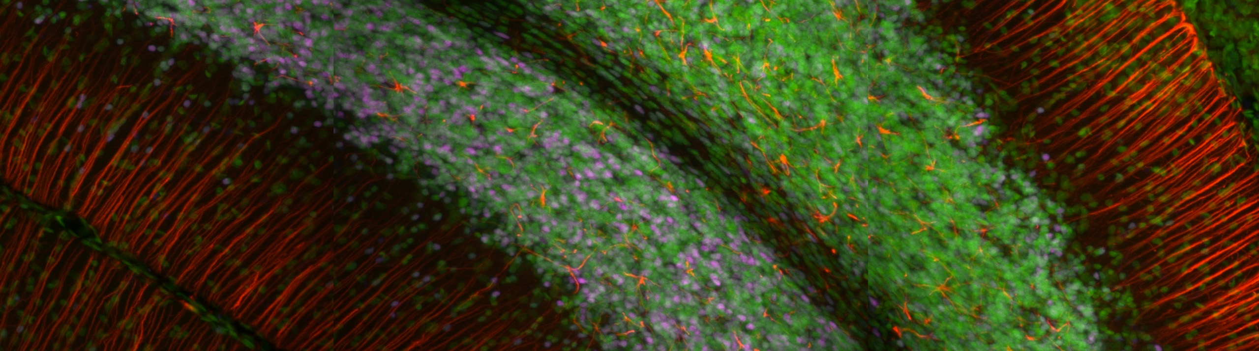
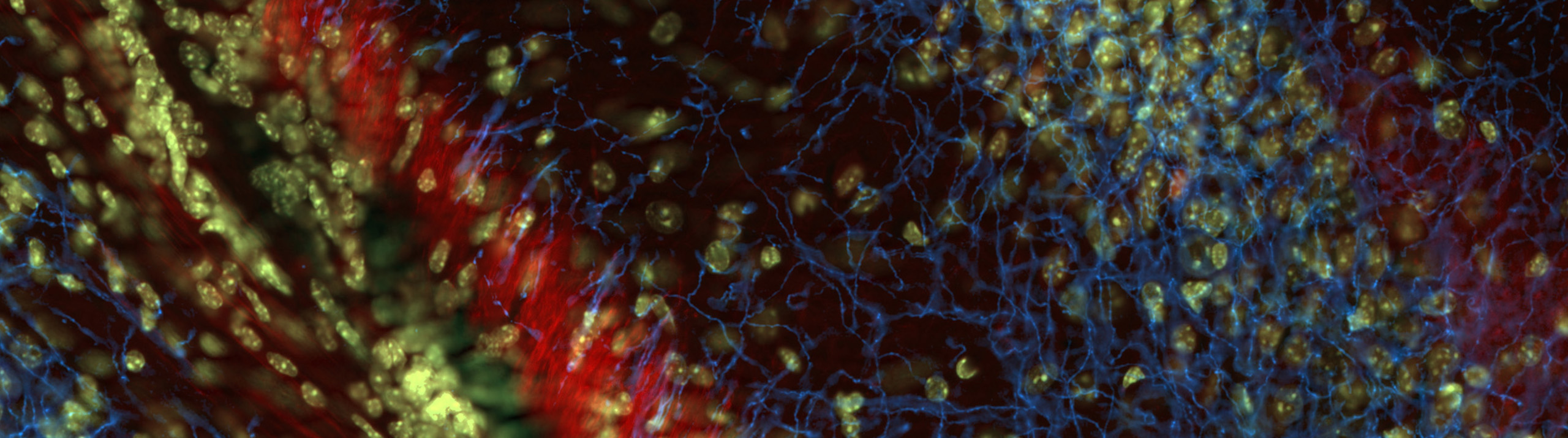
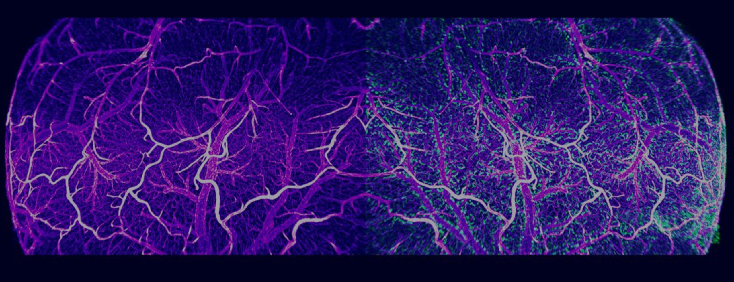
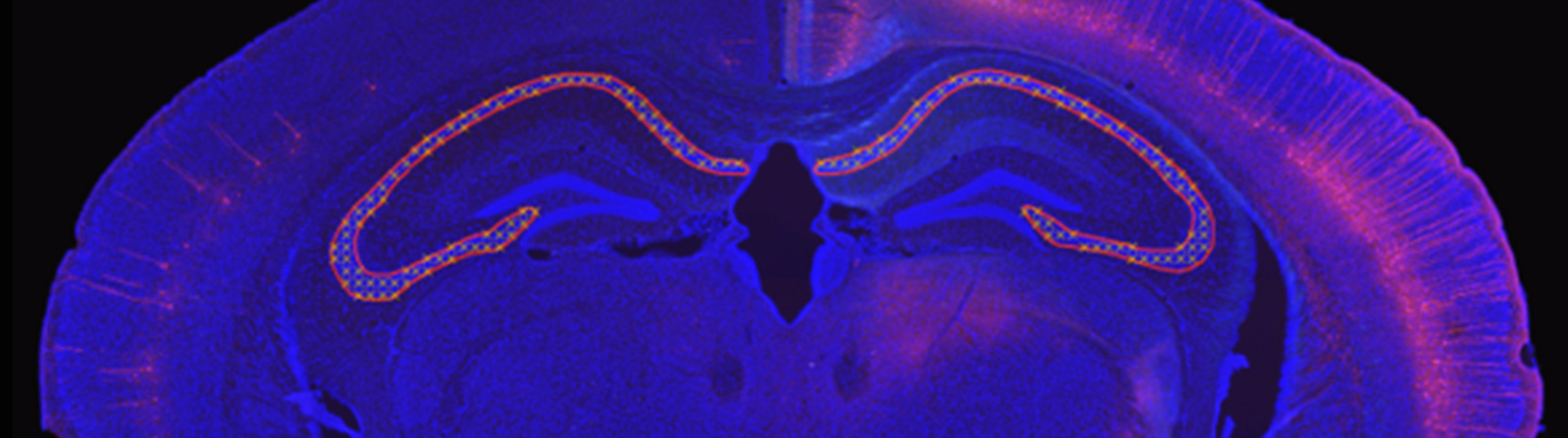
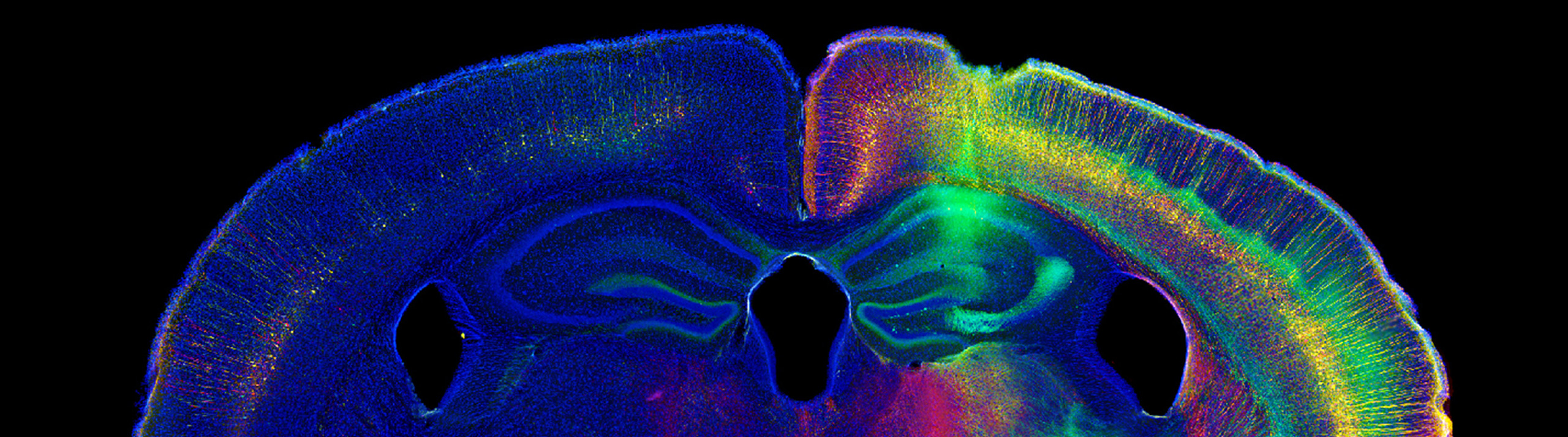
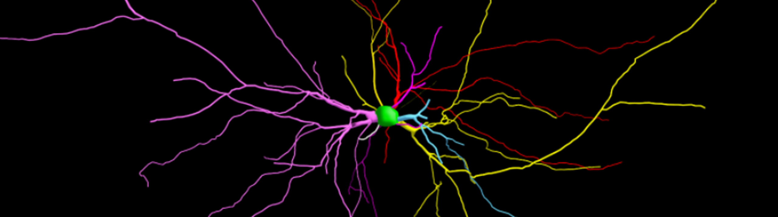
MBF Bioscience is now leveraging how neurons learn in order to improve neuroscience research using microscopy. By incorporating artificial neural networks into MBF Bioscience software, we’re equipping neuroscientists with tools that characterize neuronal populations with unprecedented accuracy and anatomic specificity through entire brain volumes.   In the webinar titled, “Improved detection of c-fos labeled and pyramidal neurons using deep machine learning in NeuroInfo,” Dr. Gerfen, joined by...

Read More

Our state-of-the-art software for performing unbiased stereology is getting an upgrade. Faster, stronger, and better, has been a theme for us lately, as we improve our products across the board, but one place where this is especially striking is in Stereo Investigator.   Set for release this spring, the new and improved Stereo Investigator will include a new imaging engine, display engine, automatic camera alignment, automatic lens calibration, the double disector, and live video zooming.     “I’m...

Read More

For Immediate Release  Williston, VT (December 10, 2020) — The ability to image large, intact biological specimens is about to get a whole lot better. Developed in collaboration with Columbia University, MBF Bioscience’s revolutionary light sheet theta microscope system, ClearScope®, has the ability to image whole tissue quickly and gently, in high resolution 3D.     The ClearScope system features a unique, two-axis scanning mode that goes beyond the capabilities of typical...

Read More
This is an image of a full Light Sheet Microscope called ClearScope

For Immediate Release: Williston, VT (February 04, 2020) — MBF Bioscience’s revolutionary light sheet microscope system, ClearScope, sets a new standard for microscopic imaging.   The new decade is poised to bring about incredible scientific innovations, and MBF Bioscience is leading the charge in 2020 with the creation of the “light sheet theta microscope” system, ClearScope.   MBF Bioscience secured exclusive license from Columbia University to develop the light...

Read More

Williston, VT – September 5, 2018 – Researchers studying microvascular networks and vessels have a groundbreaking new software application to facilitate their work. Developed by MBF Bioscience, Vesselucida®360 automatically reconstructs and analyzes microvascular networks in 3D.   Specifically designed to recognize the intricacies of the vascular system, Vesselucida features sophisticated algorithms that quickly and accurately create 3D reconstructions of images and tissue specimens. Built-in analyses provide data...

Read More

Williston, VT – June 29, 2018 – MBF Bioscience is pleased to announce the launch of the Whole Slide Edition of Stereo Investigator. This is a new version of our renown Stereo Investigator software, designed especially for conducting stereology on images from slide scanners.   Featuring a streamlined, user-friendly interface, this new product includes a variety of probes for quantifying number, length, volume, and surface area of...

Read More

Free resource for educators and researchers features thousands of downloadable histology slides   Educators and researchers around the world now have free access to a database of whole slide images (also known as virtual slides) for histology and pathology. Featuring thousands of virtual slides contributed by 15 universities, The Virtual Microscopy Database, VMD, (http://www.virtualmicroscopydatabase.org/) is an online resource that allows educators to view and download virtual images...

Read More

[caption id="attachment_6839" align="aligncenter" width="632"] Image Courtesy: Bob Jacobs, Ph.D. , Colorado College[/caption]   With the release of its new version on November 28, NeuroMorpho.org adds 9,987 new images to its archive, bringing its impressive collection of digitally reconstructed neurons to 80,012.   Scientists used MBF Bioscience’s software, Neurolucida and Neurolucida 360, to reconstruct the majority of these cells. In fact, 64 times more neurons were reconstructed with MBF Bioscience...

Read More

[caption id="attachment_6830" align="aligncenter" width="632"] An experimental coronal mouse brain section automatically aligned to the Allen Mouse Brain Reference Atlas[/caption]   Analyzing cellular populations within specific anatomies in brain images requires expertise in both neuroanatomy and cellular identification. This typically involves a scientist comparing experimental images with a reference atlas and manually delineating anatomical regions and marking cell populations within. NeuroInfo®, a revolutionary new technology from MBF Bioscience,...

Read More

  DNA damage occurs in human cells at a constant rate. These cells are usually able to repair themselves, but sometimes deficiencies in certain genes cause the repair process to shut down. When damaged DNA isn't fixed, mutations can occur that cause accelerated aging or cancerous tumors to form (Hoeijmakers, 2009). Scientists at Erasmus University Medical Center in Rotterdam have found a way to slow down...

Read More