
In Vivo Two-Photon Synaptic Imaging with ScanImage — New Findings Challenge the Hebbian Theory of Plasticity
Most neuroscientists are familiar with the saying “cells that fire together wire together,” which is often used to summarize the Hebbian theory of synaptic plasticity—first put forth in Donald Hebb’s book The Organization of Behavior in 1949. The theory describes how coincident activity between pre- and post-synaptic cells can shape synaptic strength. Verified decades later, the theory has since become accepted within the neuroscientific community.
However, a new study published in Nature, challenges Hebb’s theory. Conducted by researchers at the Max Planck Florida Institute for Neuroscience, the authors demonstrate that synapse size is not correlated with the response similarity between input and output and suggest that neural response properties reflect the total number of active synapses, both weak and strong.
Combining in vivo two-photon synaptic imaging and electron microscopy, the scientists measured the strength of excitatory inputs contacting pyramidal neurons in the primary visual cortex of the ferret brain. “These findings were made possible by the RMR scanner and ScanImage software, which allowed flexible and simultaneous in vivo recording of a large number of synapses,” explained Dr. Scholl. “In particular, this technology allowed sampling from long dendritic segments, recording multiple dendritic locations simultaneously, at synaptic resolution.”
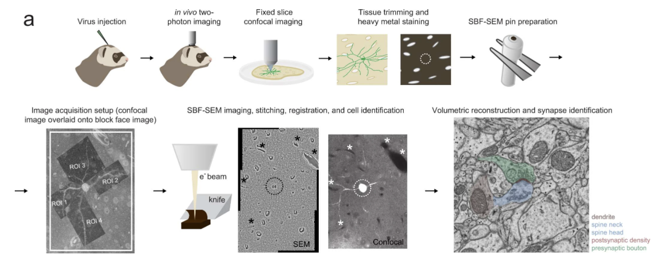
a, In vivo two-photon synaptic imaging of L2/3 cortical neurons in ferret visual cortex expressing GCaMP6s is performed under visual stimulation.
Using electron microscopy reconstruction of individual synapses as a metric of strength, we find no evidence that strong synapses have a predominant role in the selectivity of cortical neuron responses to visual stimuli. Instead, selectivity appears to arise from the total number of synapses activated by different stimuli. Moreover, spatial clustering of co-active inputs appears to be reserved for weaker synapses, enhancing the contribution of weak synapses to somatic responses. Our results challenge the role of Hebbian mechanisms in shaping neuronal selectivity in cortical circuits, and suggest that selectivity reflects the co-activation of large populations of presynaptic neurons with similar properties and a mixture of strengths. (Scholl, et.al. 2021)
For more about how ScanImage can help with your research, visit our website.
Citation:
Scholl, B., Thomas, C.I., Ryan, M.A. et al. Cortical response selectivity derives from strength in numbers of synapses. Nature 590, 111–114 (2021). https://doi.org/10.1038/s41586-020-03044-3