
Mini2P miniature microscope and ScanImage
ScanImage software from MBF Bioscience, along with the accompanying vDAQ acquisition and control card with analog to digital, digital to analog, breakout board, is built to control many combinations of hardware in order to carry out in-vivo imaging on a cellular scale. This makes it possible to observe the neural activities, such as those indicated by calcium concentration or voltage changes, of specific neuronal types identified with fluorescent labels in the intact and behaving animal. The developers of the innovative Mini2P microscope (Zong, et al. 2022) at the Kavli Institute, chose ScanImage and vDAQ as the software and hardware to control the Mini2P microscope. MBF’s software engineers worked with them to customize ScanImage and to support the hardware in the Mini2P. The hardware for the Mini2P can be grouped as components in the resource configuration dialog box of ScanImage for easy access and implementation.
The radical new advantage of the Mini2P microscope is its light weight and the flexibility of its laser-cable. With heavier microscopes, for instance when used for imaging neurons in the striatum (Maltese et al. 2021), animals are restricted to less active behaviors such as walking on a treadmill, but with the extremely light weight Mini2P, in-vivo imaging, for instance of ‘place-cells’ in the hippocampus, can be done on animals engaging in much more active behaviors, such as finding their way through a maze (Zong et al.).
ScanImage software gives efficient control of the Mirrorcle (Mirrorcle Technologies, Inc., Richmond CA) MEM scanner for XY imaging, the electronically-tunable µTlens for focusing, and laser beam power. ScanImage receives the resulting data from photo-multiplier-tubes (PMTs) and assembles images of functioning neurons. The input and output signals for this in-vivo imaging are controlled with and exchanged between ScanImage and the hardware via the VDAQ card and breakout board.
Proof of concept is shown in, ‘Large-scale two-photon calcium imaging in freely moving mice,’ by Zong et al., published this year: ScanImage … fully supports the hardware control and data acquisition of MINI2P. Following the wiring illustration and the operation manual in Methods S1, Section 9, the system can be run directly without further modification. (Zong et al., Control and Acquisition, p. e9)
The just three-gram 2P miniscope with its flexible fiber laser cable attached was shown to be light enough for active behavorial experiments. Three-dimensional imaging of fluorescence indicating cellular calcium concentration in visual cortex, hippocampus archicortex, and hippocampus was done at 7.5 Hz. The mice were so unencumbered as to be considered freely moving, and ‘place-cells’ in the hippocampus were seen to undergo changes in calcium concentration correlated with the animals’ position in the maze.
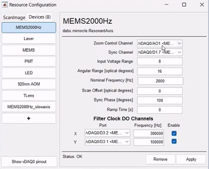
How does ScanImage accomplish control and acquisition? The mirrorcle resonant scanner driver is intended for use with a MEMS mirror device. This driver enables one of the axes, the X axis, of the mirror to be used in resonant scanning mode. To use the second axis, the Y axis, of the mirror, an Analog Galvo device is added to ScanImage. ScanImage takes the information about the fast X axis mirror position and uses it to calculate and send the signal for the relatively slow Y axis mirror. Two analog outputs of vDAQ send the MEMS scanning control signal (fast axial and slow axial) to the mirror device. A third analog output sends the control signal to the µTlens driver (Thorlabs, Newton, NJ). A fourth analog output sends the laser power control signal to the laser controller. The laser source was a compact, single-wavelength, fiber-based femtosecond laser (FemtoFiber Ultra 920, Toptica, Munich, Germany). For acquisition, the signals from two-channel PMTs are connected to two high-speed (125 MHz) analog inputs of the vDAQ card. A maximum of 4 channels can be acquired simultaneously. ScanImage organizes the PMT data to form the images.
The process of imaging functioning neurons in freely behaving animals is incredibly powerful, but also exceedingly complex. ScanImage software can simplify these procedures and make the control of in-vivo image acquisition easily executable.
To learn more about how ScanImage controls the Mini2P, click here to see the workshop given by ScanImage product manager, Mitchell Sandoe.
Reference:
Maltese, Marta, Jeffrey R March, Alexander G Bashaw, Nicolas X Tritsch, 2021, Dopamine differentially modulates the size of projection neuron ensembles in the intact and dopamine-depleted striatum. https://doi.org/10.7554/eLife.68041
Zong, Weijian, Horst A Obenhaus , Emilie R Skytøen, Hanna Eneqvist, Nienke L de Jong, Ruben Vale, Marina R Jorge, May-Britt Moser, Edvard I Moser, 2022, Large-scale two-photon calcium imaging in freely moving mice. Cell, 185(7):1240-1256.e30. doi: 10.1016/j.cell.2022.02.017. Epub 2022 Mar 18.