Additional Subject Matter

MBF Bioscience >  Blog > Additional Subject Matter (Page 2)

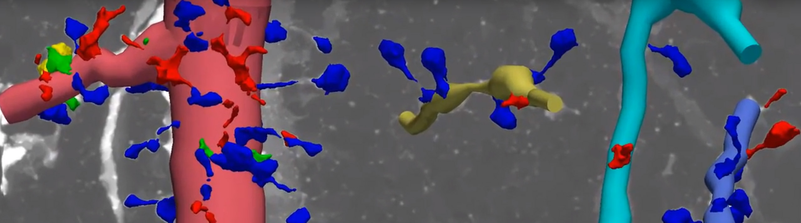
We are pleased to announce that the International Neuroinformatics Coordinating Facility (INCF) has endorsed the MBF Bioscience neuromorphological file format as a standard.   The file format is used in our products for neuroscience research for important applications such as digital neuron tracing, brain mapping and stereological analyses. MBF Bioscience products, including Neurolucida, Neurolucida 360, Stereo Investigator, Vesselucida 360, and NeuroInfo use this neuromorphological file format.   This file...

Read More

When we flex our thumb or point our finger, axons carry impulses from the brain to neurons in the spinal cord, which send messages to the muscles in our hands. In an important study in 1983, Jenny and Inukai at the Washington University School of Medicine reported the organizational patterns of those finger movement motoneuron columns in the primate spinal cord. Now, nearly 40 years...

Read More

Obesity has reached epidemic levels worldwide. It is a major cause of deadly illnesses, such as diabetes, cardiovascular disease, and some cancers, and has been linked to reduced quality of life and poor mental health outcomes. Efforts to combat this epidemic have so far failed to have an appreciable impact.   Seeking novel targets with the potential to move the needle on obesity-linked cardiometabolic disorders, a research...

Read More

MBF Bioscience proudly continues its commitment to neuroscience advances.   A century ago, when neuroscientists were asked about the characteristic properties of a nerve cell (neuron), they answered that neurons are cells in the central nervous system (CNS) characterized by a soma (cell body), processes that receive information from other neurons (dendrites), and a process that sends information to other neurons (axon). This answer has not changed...

Read More
High Resolution scan of brain

Researchers studying structure and function in rat brain can now use NeuroInfo to analyze and register their brain volumes to the Waxholm Rat Brain Atlas Version 4—an open access volumetric atlas of the Sprague Dawley rat brain.   “NeuroInfo already includes extensive analysis capabilities for mouse brain research by standardizing measurements on brain volumes to the Allen Mouse Brain Atlas. The inclusion of a rat atlas in...

Read More

Dear MBF scientific community, As another year comes to a close, we at MBF are thinking of all of you.   We’ll always remember 2021 as the “recalibration year” of the pandemic — vaccines, reopenings, variants, lockdowns coming and going, conferences being held virtually or canceled altogether, social distancing from each other and all of you.   As they say, you don’t know what you miss until it’s gone. And...

Read More

Our health depends on the ability of blood vessels to deliver nutrients and remove metabolic byproducts from organs and muscle systems. But what happens to this delicately balanced process after traumatic injury? Scientists generally understand that skeletal muscles can regenerate, but little is known about how this happens at the level of our microvasculature.   [caption id="attachment_7659" align="aligncenter" width="699"] Representative maps of resistance networks from feed artery...

Read More

I hope this missive finds all of you healthy and happy. The pandemic continues to interrupt our daily lives, and our ways of working and doing business. I was particularly disappointed about the cancelation of the in-person portion of this year’s Society for Neuroscience conference. I was so looking forward to seeing so many of you in person, there’s just no substitute for being connected...

Read More

Most neuroscientists are familiar with the saying “cells that fire together wire together,” which is often used to summarize the Hebbian theory of synaptic plasticity—first put forth in Donald Hebb’s book The Organization of Behavior in 1949. The theory describes how coincident activity between pre- and post-synaptic cells can shape synaptic strength. Verified decades later, the theory has since become accepted within the neuroscientific community.   However,...

Read More

What if, instead of trying to zoom in on the nanoscale structures of the brain, we made them bigger? Dr. Edward Boyden and his team at MIT are doing just that. The process, known as expansion microscopy (ExM), physically enlarges brain tissue so that even extremely small molecular structures are viewable on a conventional light microscope.   In his 2016 TED Talk, Dr. Boyden says: “Can we make...

Read More