MBF Bioscience Release ClearScope, Revolutionary Light Sheet Microscope for Imaging Large and Small Cleared Tissue Specimens
For Immediate Release
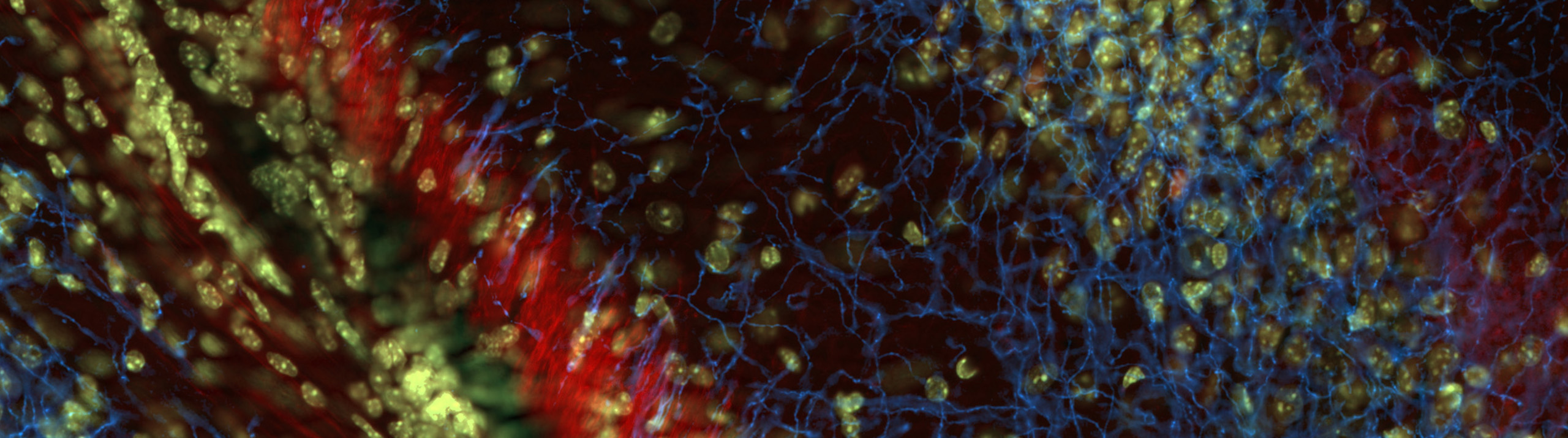
Williston, VT (December 10, 2020) — The ability to image large, intact biological specimens is about to get a whole lot better. Developed in collaboration with Columbia University, MBF Bioscience’s revolutionary light sheet theta microscope system, ClearScope®, has the ability to image whole tissue quickly and gently, in high resolution 3D.
The ClearScope system features a unique, two-axis scanning mode that goes beyond the capabilities of typical light sheet microscopy. The patent pending design ensures the specimen is constantly illuminated using the thinnest section of the light sheet optimizing axial resolution. Shadow effects and uneven illumination, which hindered analysis in the past, are eliminated by the new technology, so that even the smallest subcellular structures within the tissue can be visualized with precision.
Compatible with a range of tissue clearing techniques, including CLARITY, iDISCO, uDISCO, SeeDB and Sca/e, ClearScope has the ability to capture the details of very thick, very large specimens, such as intact tissue from human and primate brains. President and co-founder of MBF Bioscience, Jack Glaser says, “With low photo-bleaching, fast imaging speed, and high image quality, ClearScope is the most advanced tissue-imaging system in development today, and is set to revolutionize brain research.”
Designed to work with a wide range of cleared specimens, the system is completely customizable and easily adapted to address the specific needs of researchers across a variety of disciplines.
If you are interested in learning more about MBF Bioscience’s advanced new system for imaging large tissue specimens, visit: https://www.mbfbioscience.com/clearscope
###
About MBF Bioscience: MBF Bioscience integrates the world’s leading microscope systems with our revolutionary quantitative imaging and visualization software to accelerate research in the fields of: stereology, neuron and microvasculature reconstruction, vascular analysis, worm tracking, brain mapping and big image data management in medical research and education.
Since 1988, MBF Bioscience has forged a rich history of creating innovative products to empower biological researchers with the quantitative analysis tools they need to obtain accurate, unbiased results. With offices in North America, Europe, Japan, and China, MBF Bioscience has helped researchers across the globe publish over 15,000 peer-reviewed papers in peer reviewed journals. MBF Bioscience partners with the NIH and distinguished scientists across the world to continue their commitment to neuroscience research with their software technology, and also in the fields of stem cells, pulmonology, oncology, and toxicology. For more information visit www.mbfbioscience.com or follow MBF Bioscience on Facebook, Twitter, and LinkedIn.
###
Media Contact:
Pasang Sherpa
Marketing Manager
1-802-288-9290
pasang@mbfbioscience.com