Magnified: C. Elegans Captured with Lumenera CMOS Camera
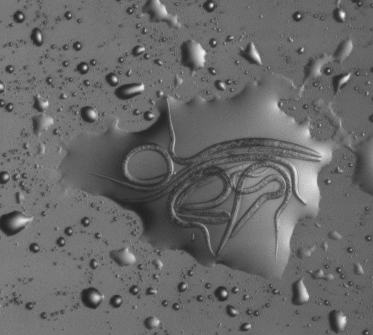
MBF Bioscience Vice President of Research Jeff Sprenger captured this exceptional image of c.elegans worms while testing out the Lumenera CCD Lu135M digital camera. He was working with our new WormLab software, which is set for release next week. Here Jeff shares the details about how he captured the image:
This image was captured on our experimental WormLab setup, using a macro-imaging stand and setup devised here at MBF Bioscience. The c. elegans worms are trapped in a drop of liquid on an agar plate (60mm petri dish). We’re testing a Lumenera CCD Lu135M digital camera, using an exposure time of 120 ms and gain of 2.0X with no gamma adjustment for this image. The lens is a Canon Macro zoom MPE-65mm, with c-mount adapter. The light source is an MBF Bioscience LED light plate, with a custom diaphragm and polarizing filter to increase contrast.
WormLab, software for tracking crawling microscopic worms, is set for official release next week. C. elegans (caenorhabditis elegans) are commonly used by geneticists and neuroscientist to study life span, regulation of metabolism, behavior and development.
Learn more about WormLab on our website.
For the latest news about MBF Bioscience and our customers, fan us on Facebook and follow us on Twitter.