Neurolucida Helps Scientists Discover that Gorillas are Relevant in the Study of Alzheimer’s Disease
Humans and gorillas are approximately 98% identical on a genetic level, however there is little published research exploring Alzheimer’s disease pathology in gorillas. A new paper reports that gorillas display similarities in advanced age to humans ̶ including the presence of Alzheimer’s disease precursors like amyloid-beta (Aβ) plaques and tau lesions.
The study, published in the Journal of Comparative Neurology, provides evidence of Alzheimer’s disease precursors in the western lowland gorilla. Their findings broaden the scientific community’s understanding of the aging brain of some our closest living relatives and offer new insights for Alzheimer’s disease research.
“As great apes and humans share numerous anatomical and physiological similarities due to their close genetic ancestry, the knowledge of interspecies differences in AD-like pathology could provide clues to the mechanisms underlying AD pathology in humans,” the authors say in their paper.
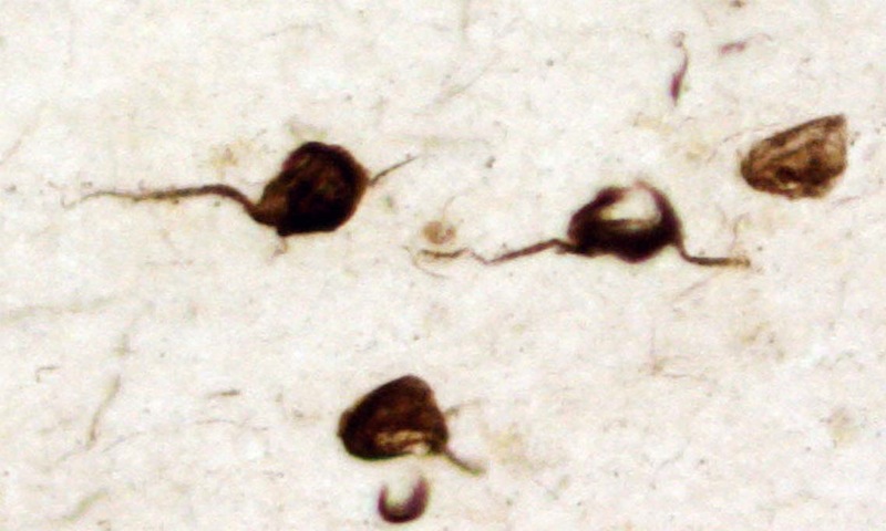
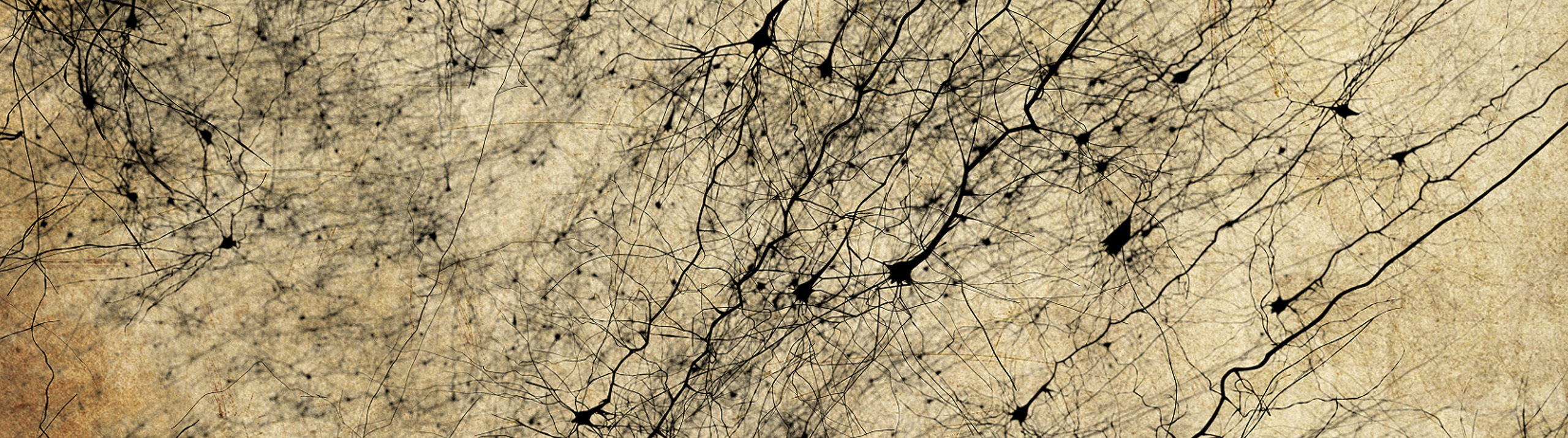
Cortical neurons containing tau, termed neurofibrillary tangle, seen in the human brain with Alzheimer’s disease. The researchers used Neurolucida to chart similar lesions in the gorilla brain.
The researchers studied neocortical and hippocampal specimens from western lowland gorillas provided by the Great Ape Aging Project, an organization dedicated to the advancement of knowledge about great apes. Formerly housed at zoos and aquariums around the United States, the gorillas all died of natural causes at ages ranging from 13 to 55-years-old.
Their qualitative analyses of immunohistochemically stained sections of the apes’ frontal cortices and hippocampi revealed an age-related increase in Aβ plaques and Aβ-positive blood vessels, with “many more” APP/Aβ-positive plaques in the cortex of the oldest gorilla – the 55-year-old female, compared with the 50-year-old female. They also saw higher levels of Aβ plaques in the neocortex and hippocampus of females, while male gorillas had more Aβ-positive blood vessels. Also interesting were the differences at varying cortical levels. The researchers saw compact, strongly labeled plaques in the subgranular layers (I-III) and paler, larger plaques in the infragranular layers (V-VI), according to the paper.
Then, the researchers used Neurolucida to map APP/Aβ-immunoreactive plaques and Alz50-immunoreactive neurons in the neocortex and hippocampus of the 55-year-old female. Alz50 is a stain for a phosphorylated epitope of tau protein. Tau is the main component of neurofibrillary tangles.
“Neurolucida allowed us to chart labeled images in a timely and efficient manner,” Dr. Elliot Mufson, a lead author of the study said.
In summary, this research paper reports the first evidence of age-related Aβ plaques in gorillas, and the first evidence of tau-positive profiles in gorillas. These findings show that gorillas are relevant in the study of Alzheimer’s disease.
Perez, S. E., Raghanti, M. A., Hof, P. R., Kramer, L., Ikonomovic, M. D., Lacor, P. N., Erwin, J.M., Sherwood, C.C., Mufson, E. J. (2013). Alzheimer’s disease pathology in the neocortex and hippocampus of the western lowland gorilla (Gorilla gorilla gorilla). Journal of Comparative Neurology, doi: 10.1002/cne.23428