Florida Researchers Study Traumatic Brain Injury With Stereo Investigator
If a head gets hit hard enough, the trauma occurs instantly. Neurons die, the brain swells as microglia cells rush to the damaged area, and the protective armor known as the blood brain barrier might even rupture. But it doesn’t end there. Long term effects include cognitive impairment, loss of sensory processing, and susceptibility to neurodegenerative diseases like Alzheimer’s.
Researchers at the University of South Florida say patients suffering from chronic Traumatic Brain Injury (TBI) experience a “cascade of events” marked by long-term neuroinflammation, cell loss, and impaired cell proliferation that may manifest over time.
“While TBI is generally considered an acute injury, a chronic secondary cell death perturbation (i.e., neuroinflammation) and a diminished endogenous repair mechanism (i.e., cell proliferation) accompany the disease pathology over long-term,” the authors say in their paper published this month in PLOS ONE.
The scientists used unbiased stereology to analyze activated microglia cells, cell proliferation, and differentiation into immature neurons in several regions of the brains of rats which had experienced TBI eight weeks prior.
They used Stereo Investigator with the Cavalieri estimator probe and the optical fractionator probe to estimate the quantity and volume of stained cells in the cortex, striatum, thalamus, fornix, cerebral peduncle, and corpus callosum, as well as the subgranular zone and the subventricular zone in both hemispheres of the brain.
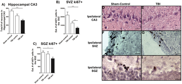
Figure 3 from “Hippocampal CA3 cell loss and downregulation of cell proliferation.”
Eight weeks after the TBI occurred, the researchers found an increased level of active microglia cells at the direct site of the TBI as well as surrounding regions. They also report a decrease in hippocampal neurons, and low levels of cell proliferation in the neurogenic niches.
“Our overarching theme advances the concept that a massive neuroinflammation after TBI represents a second wave of cell death that impairs the proliferative capacity of cells, and impedes the regenerative capacity of neurogenesis in chronic TBI,” the authors say in their paper.
They go on to suggest a “multi-pronged treatment targeting inflammatory and cell proliferative pathways” may help alleviate the pathological effects of chronic TBI.
Read the full paper “Long-Term Up-regulation of Inflammation and Suppression of Cell Proliferation in the Brain of Adult Rats Exposed to Traumatic Brain Injury Using the Controlled Cortical Impact Model” on PLOS ONE.
Source: Acosta S.A., Tajiri N., Shinozuka K., Ishikawa H., Grimmig B., et al. (2013). Long-Term Up-regulation of Inflammation and Suppression of Cell Proliferation in the Brain of Adult Rats Exposed to Traumatic Brain Injury Using the Controlled Cortical Impact Model. PLoS ONE 8(1): e53376. doi:10.1371/journal.pone.0053376