3D Reconstruction

MBF Bioscience >  Blog > Additional Subject Matter  > 3D Reconstruction (Page 4)

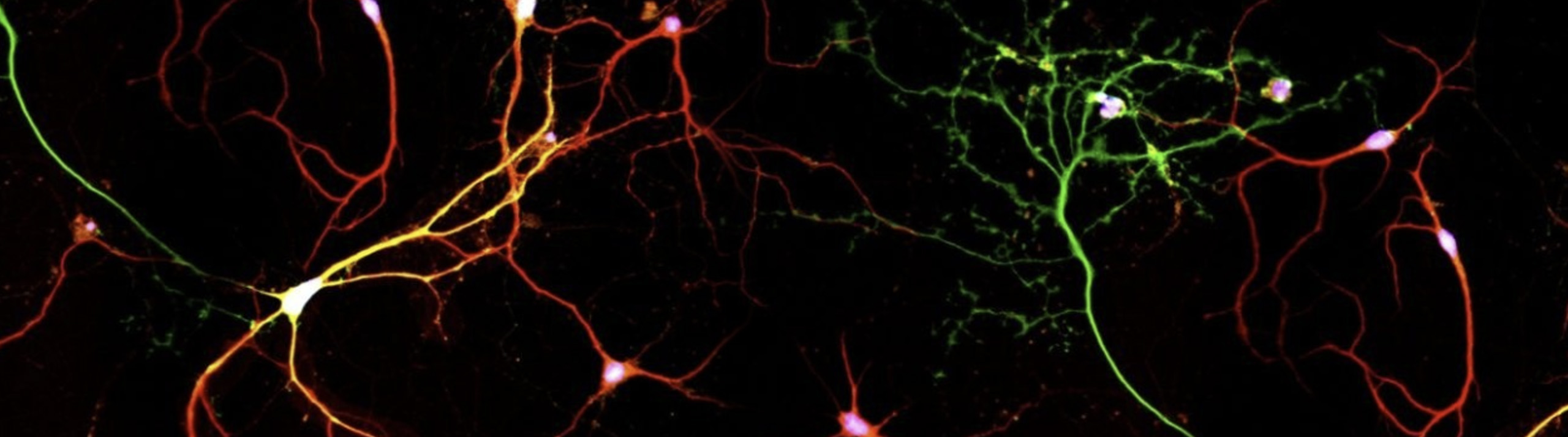
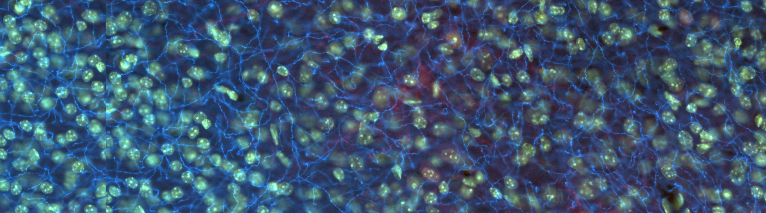
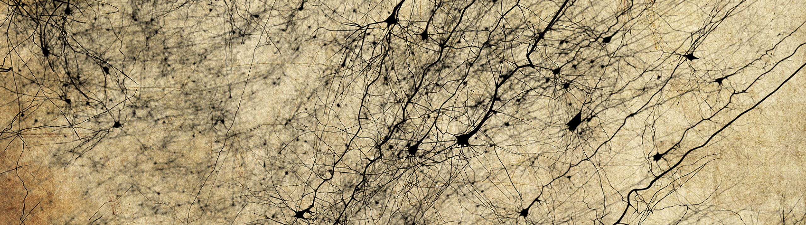
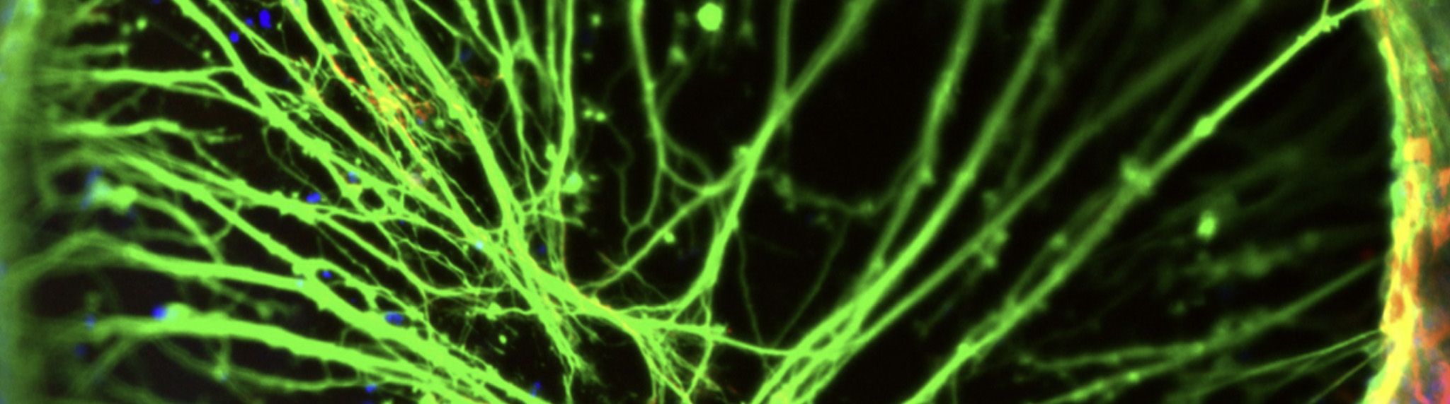
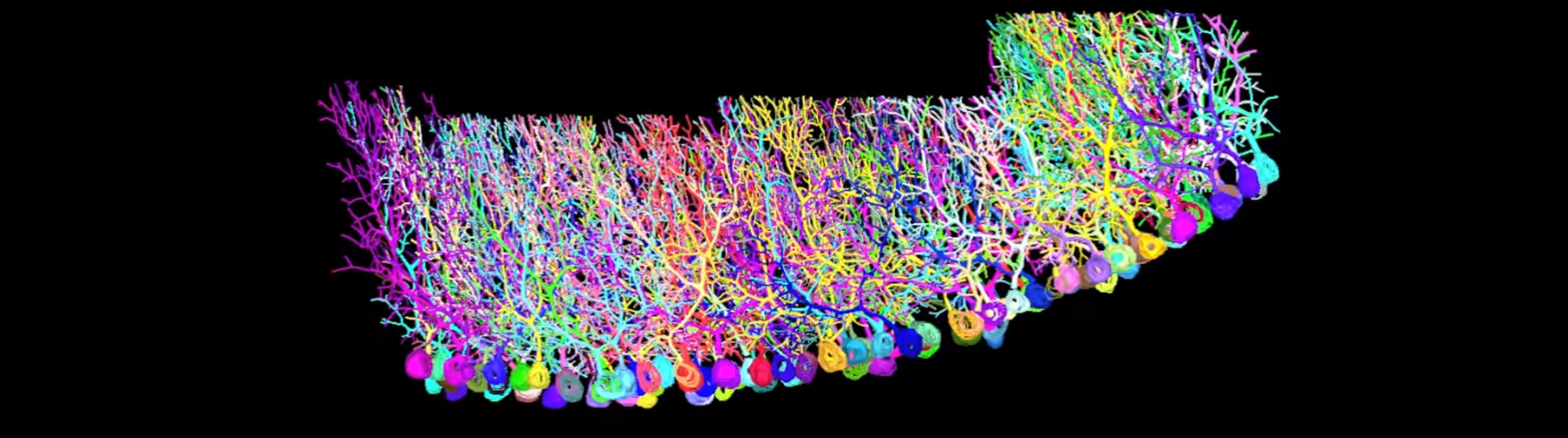
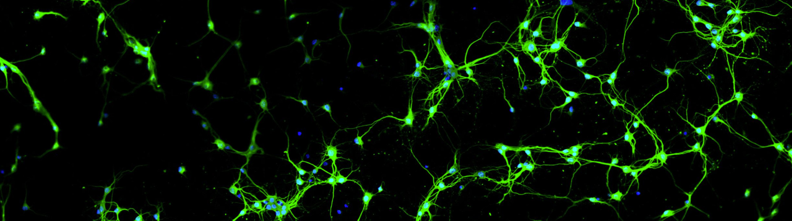
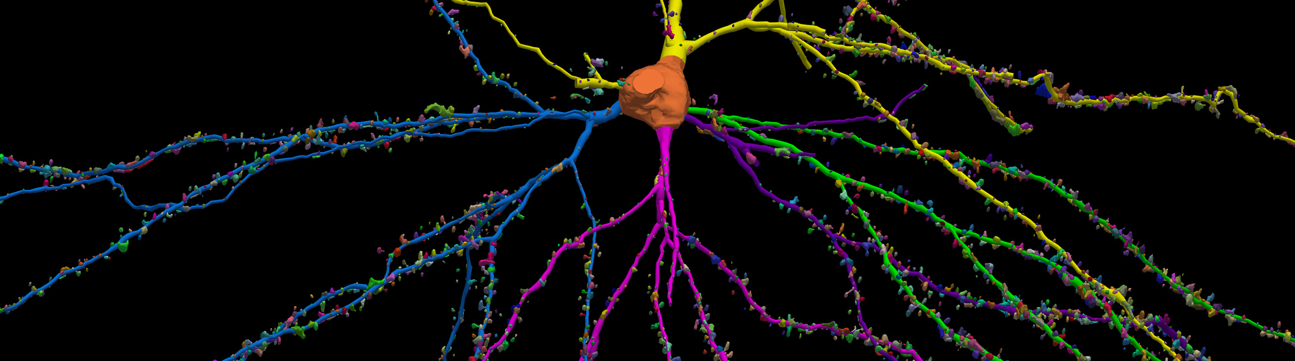
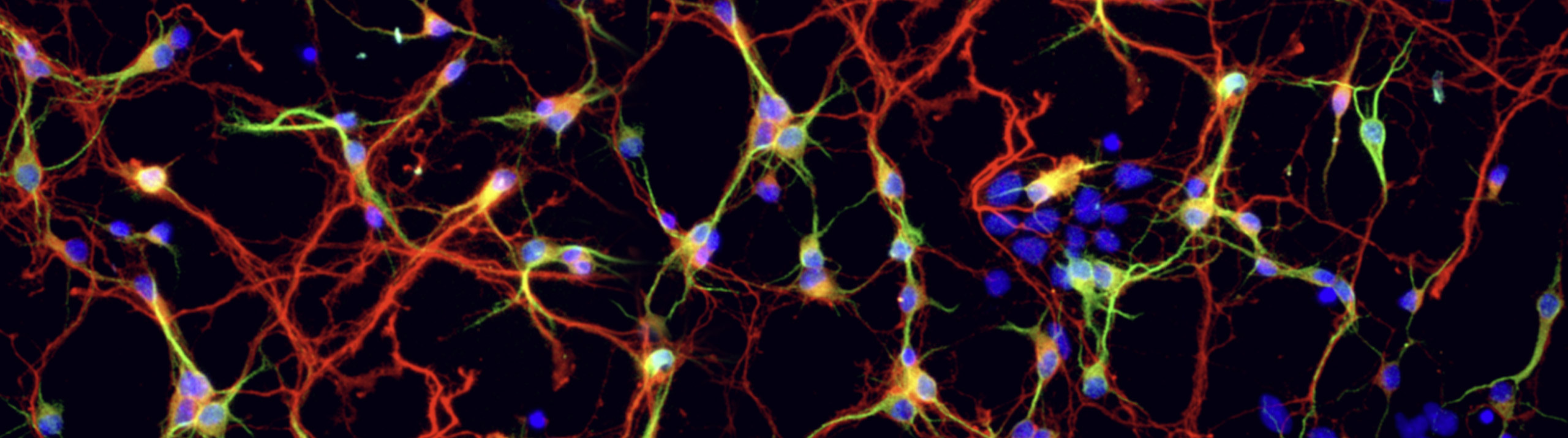
When it comes to preferred neuron reconstruction systems, Neurolucida “dominated the last decade” according to a paper published earlier this year in Frontiers in Neuroscience. The paper, “Digital reconstructions of neuronal morphology: three decades of research trends" (Halavi et al, 2012), offers an overview of the history of digital neuron reconstruction and presents research trends on specific animal species, brain regions, neuron types, and experimental approaches. Beginning...

Read More

Blue Brain Project researchers have hit an important milestone in their quest to create a virtual model of the human brain. They figured out how to accurately predict the location of synapses in the neocortex; and Neurolucida played an important part.   In a paper published last week in PNAS, the research team led by Dr. Henry Markram at the Brain Mind Institute at the Ecole Polytechnique Fédérale...

Read More

During a chicken embryo's twenty-one days of incubation, its eyes develop in astonishing ways. Muscles form, neurons branch, innervation occurs. Researchers at Dr. Rae Nishi's lab at the University of Vermont, including two MBF Bioscience staff scientists Julie Simpson, Ph.D. and Julie Keefe, M.S. are studying the development of a chicken embryo's nervous system. Their specific focus is on the behavior of neurons in the...

Read More

  When an adult rat learns new things about its physical environment, the newborn neurons in its brain change – dendrites branch, spines increase, soma grows. But what about mature neurons? Might they also undergo structural changes in response to learning? “Yes,” say scientists at the National Institute of Health and Medical Research and the University of Bordeaux, in Bordeaux, France.   Led by Drs. Valérie Lemaire, Sophie...

Read More

A rat uses its whiskers to get information about its environment. As it scurries along the subway tracks, or burrows into a dumpster, its whiskers send signals to ascending parts of its brain that let it know for example, whether it is safe to jump over that gap or not.   Scientists at the Max Planck Florida Institute are studying the functional responses of neurons in the...

Read More

Using Neurolucida, microscopy, and mice genetically engineered to express a random amount of red, yellow, and blue fluorescent proteins, Okinawa Institute of Science and Technology researcher Hermina Nedelescu has created a fascinating and hypnotic movie of neurons. Nedelescu and colleagues at the Institute's Computational Neuroscience Unit used Neurolucida and its Virtual Tissue 3D Extension Module and Montaging tools to acquire and stitch together multiple images of Purkinje cells—large neurons  that...

Read More

A willowy pair of pyramidal cells engage in an intricate dance with a dense mass of basket cells on the cover of the September 14, 2011 issue of the Journal of Neuroscience.   This exquisite image illustrates recent work by Columbia University researchers Dr. Adam M. Packer and Dr. Rafael Yuste, who used Neurolucida to study circuit connectivity in the mammalian neocortex.   According to the paper "Successfully filled...

Read More

No two trees are exactly alike, in the forest or in the brain. Though despite the diversity of dendritic arborizations, when it comes to branching out different types of neurons do have a couple things in common, say researchers at the National Institute for Physiological Sciences in Okazaki, Japan.   Led by longtime MBF Bioscience customer Dr. Yoshiyuki Kubota, the research team identified two organizational principles common...

Read More

When Pavlov's dogs started salivating in response to a ringing bell, something happened in their brains—a memory was encoded. Over a century later scientists are still figuring out how memories are physically represented in the brain. One lingering question has been whether or not the same set of neurons is activated when a particular memory is formed. Researchers at the Johnson Lab at the Uniformed...

Read More

What would you do with a neuron if you could activate its synapses in any combination you wanted? Tiago Branco, Beverley A. Clark and Michael Hauser created a chance to do just that (Branco, 2010). The authors, using in-vitro brain slices containing layer II/III pyramidal cells in visual or somatosensory cortex of rats, were able to excite identified spines in any order and with whatever...

Read More