
Japanese Researchers Develop New Optical Clearing Agent; Neurolucida Used For 3D Imaging in Study
A new optical clearing agent developed by scientists in Japan clears brain tissue samples with greater transparency and less time than other clearing agents, according to a paper published in Nature Neuroscience.
“Combined with two-photon microscopy, SeeDB allowed us to image fixed mouse brains at the millimeter-scale level,” say the authors, who after clearing the brain tissue with SeeDB, captured images with a multiphoton Olympus microscope, and visualized 3D reconstructions with Neurolucida.
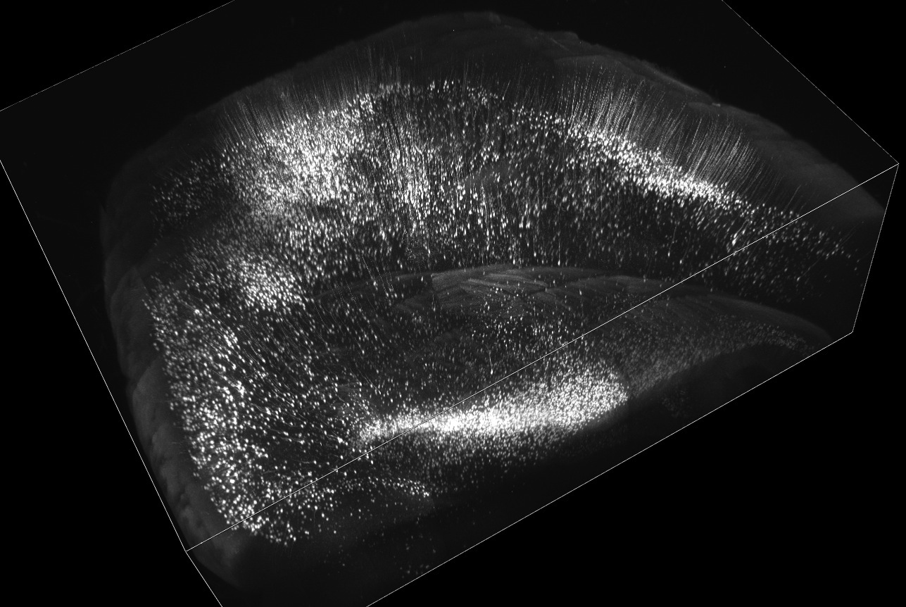
Volume rendering of mouse cerebral cortex and hippocampus. Adult Thy1-YFP-H line mouse brain was cleared with SeeDB and imaged using two-photon microscopy. Imaging area shown is 4 x 5 mm (8 x 10 tiles), 2mm thick. We could easily make a volume rendering from a large set of 3D data (in this case, 9GB two-photon data).
A solution of fructose, water, and alpha-thioglycerol, SeeDB cleared gray and white matter brain tissue samples in three days without affecting the volume or morphology of the tissue. Dendritic spines of pyramidal neurons in the cerebral cortex was one aspect of fine morphological architecture that the authors note remained intact after SeeDB treatment.
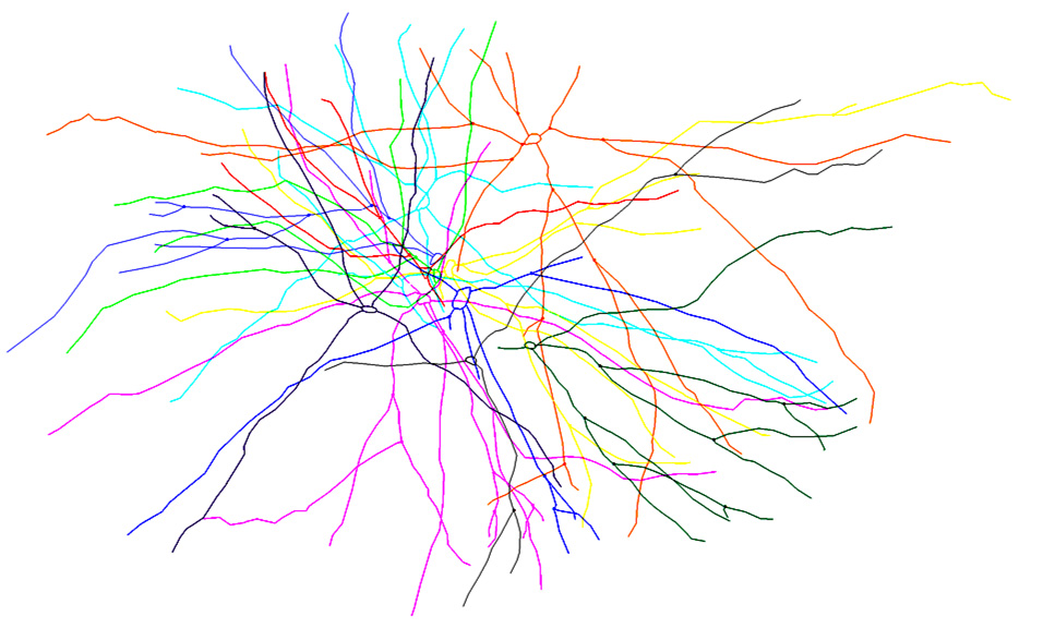
Reconstruction of lateral dendrites of sister mitral cells. Fluorescent neuronal tracer (Alexa647 dextran amine) was electroporated into a single glomerulus to label ‘sister’ mitral cells associated with a common glomerulus. After optical clearing of the olfactory bulb with SeeDB, the olfactory bulb was imaged using confocal microscopy. Lateral dendrites of labeled mitral cells were reconstructed using Neurolucida. This reconstruction was used for quantitative analysis of ‘sister’ mitral cell distribution.
Compatible with fluorescent proteins and various neuronal tracers including lipophilic dyes such as DiI, the clearing agent proved to be an effective tool for quantitative reconstructions of microcircuits in the brain. To illustrate this point, the authors examined a detailed wiring diagram in the mouse olfactory bulb. They labeled neurons with fluorescent dextran tracers, cleared the tissue with SeeDB, and using confocal microscopy, imaged the wiring diagram of the dendrites of a type of neuron known as mitral cells. To reconstruct these dendrites, the scientists analyzed confocal images and manually traced the dendrites in three-dimensional space using Neurolucida.
“SeeDB is particularly advantageous for quantitative analyses of fine neuronal morphology and microcircuits because minimal deformation artifacts occur during the clearing process,” the authors say. “Because our protocol is quick, easy, inexpensive, safe and requires no special equipment, SeeDB could prove useful for a broad range of researchers working in neuroscience and developmental biology.”
Ke, M., Fujimoto, S., Imai, T., (2013). SeeDB: a simple and morphology-preserving optical clearing agent for neuronal circuit reconstruction. Nature Neuroscience, doi:10.1038/nn.3447