
Neurolucida Helps Scientists Map Rett Syndrome’s Brain Dysfunction in Mouse Model
At first, all appears normal with the infant’s development. But one day, around her first birthday, she stops making eye contact, her babbling comes to an end, she wrings her hands, and holds her breath. The child will likely survive into adulthood, but with Rett syndrome, she will lead a life with severe disabilities.
The symptoms of this autism-related disorder are complex, and treatments are not available. At the Case Western Reserve University School of Medicine, in Cleveland, Dr. David Katz and his team of neuroscientists are researching the rare genetic disorder, which affects one in 10,000 mostly female children. Their recent study, published in the Journal of Neuroscience, describes a map of brain dysfunction in a mouse model of Rett syndrome, as well as a promising treatment with the drug ketamine.
Their first step in the study was the brain map. To create it, the researchers used Neurolucida to quantify and compare the distribution of Fos-positive cells in the brains of normal mice and those with a mutation in Mecp2, a defective gene in Rett syndrome patients. Fos is a protein whose level of expression is correlated with neuronal activity. The Katz lab used Fos labeling to detect differences in neuronal activity between different regions of the normal and mutant brain. The map – the first of its kind, showed abnormally low levels of activity in the forebrain, and abnormally high levels of activity in the brainstem in Mecp2 mutant mice.
“We discovered that many of the key regions where activity is below normal in the mutants overlap with regions that also exhibit reduced activity in human patients with classic autism. This suggests there may be common underlying mechanisms contributing to the behavioral symptoms that are shared between Rett syndrome and more common forms of autism spectrum disorders,” Dr. Katz said.
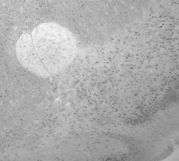
Fos labeling in the nucleus accumbens, provided by Dr. David Katz.
By pinpointing the affected brain regions, the researchers were able to determine which areas needed to be treated. Knowing from previous studies that ketamine activated neurons in the forebrain, they administered a low dose of the drug to the Rett syndrome mice. According to the study, quantitative analysis showed a return to normal levels of Fos expression in the mouse forebrain after being treated with ketamine.
“These studies provide new evidence that drug treatment can reverse abnormalities in brain function in Rett syndrome mice,” Dr. Katz said in a Case Western Reserve University press release. “They also provide new leads as to what kinds of drugs might be effective in individuals with Rett syndrome.”
Kron, M., Howell, C. J., Adams, I. T., Ransbottom, M., Christian, D., Ogier, M., & Katz, D. M. (2012). Brain activity mapping in Mecp2 mutant mice reveals functional deficits in forebrain circuits, including key nodes in the default mode network, that are reversed with ketamine treatment. The Journal of Neuroscience (40), 13860-13872. doi:10.1523/JNEUROSCI.2159-12.2012