
Drug for Treating Asthma Improves Cognitive Function in Down Syndrome Mouse Model
Researchers at the Stanford University School of Medicine have found that formoterol ̶ an FDA-approved drug for treating asthma and similar respiratory disorders ̶ improves cognitive function in mice genetically altered to exhibit symptoms of Down syndrome including cognitive disability.
Formoterol was chosen for the study because it activates β2 adrenergic receptors (β2ARs) on neurons, a task also carried out by norepinephrine, a neurotransmitter with a critical role in contextual learning. β2AR receptors play a key role in learning and memory, and are prevalent on dentate granule cells (DGCs) in the hippocampus. To limit the effects of the drug to the CNS, the authors used a βAR antagonist with no ability to cross the blood brain barrier.
Previous studies by the same group published in Science Translational Medicine has shown that locus coeruleus neurons, which are the sole suppliers of norepinephrine to the hippocampus, degenerate over time in people with Down syndrome and in the Ts65Dn mouse model of Down syndrome (Salehi et al., 2009). The current study, published in the July issue of Biological Psychiatry (Dang et al., 2013) was aimed at determining if a drug currently used by humans could restore cognitive function in a mouse model of Down syndrome.
Behavioral tests conducted in the study revealed that formoterol improved contextual learning and reduced hyperactivity in the Down syndrome mouse model. Postmortem studies focused on the dentate gyrus of the hippocampus showed that the mice treated with formoterol exhibited an increase in synaptic density and an increase in neuronal activity over controls. In addition, newly born neurons in the dentate gyrus of formoterol-treated mice were more complex than those in the untreated mouse model of Down syndrome.
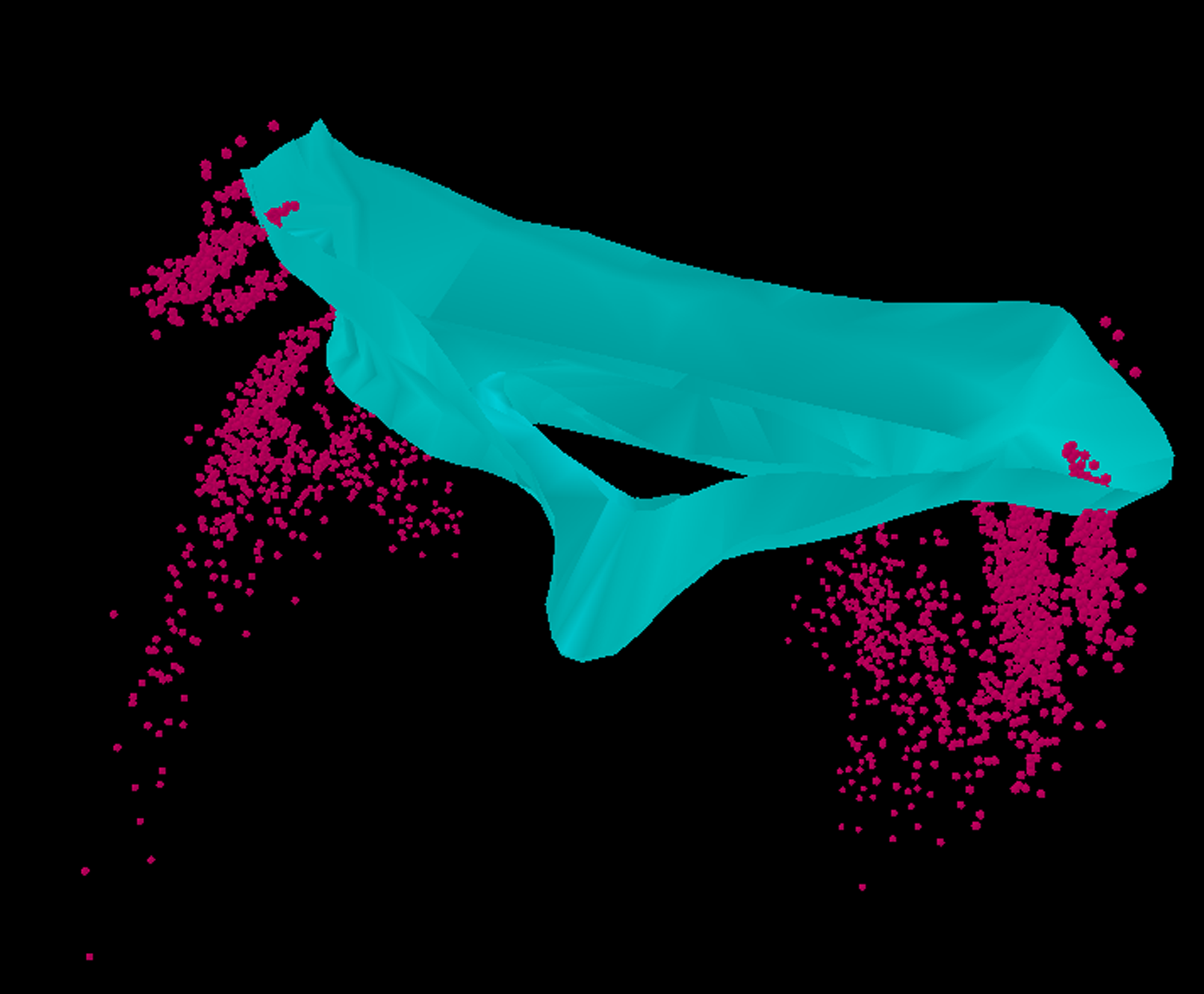 Figure 1. 3D visualization of locus coeruleus neurons (red circles) in the two corners of the 4th ventricle (green region) in rodents’ brainstem. Neurolucida was used to locate and visualize the location of immuno-labeled locus coeruleus neurons in the Ts65Dn mouse model of Down syndrome and its controls.
Researchers used Neurolucida to visualize locus coeruleus (LC) neurons (Figure 1) in the dentate gyrus (Figure 2) of the Ts65Dn mouse model of Down syndrome and found significant degeneration and atrophy of LC neurons and the dentate gyrus in the Ts65Dn mouse model. Furthermore, they studied the status of newly born neurons in the dentate gyrus (Figure 2) of formoterol-treated mice and controls. The researchers then used the Fan-In Diagram in Neurolucida to show that the formoterol-treated mice have a more complex branching pattern than the untreated mice (Figure 3).
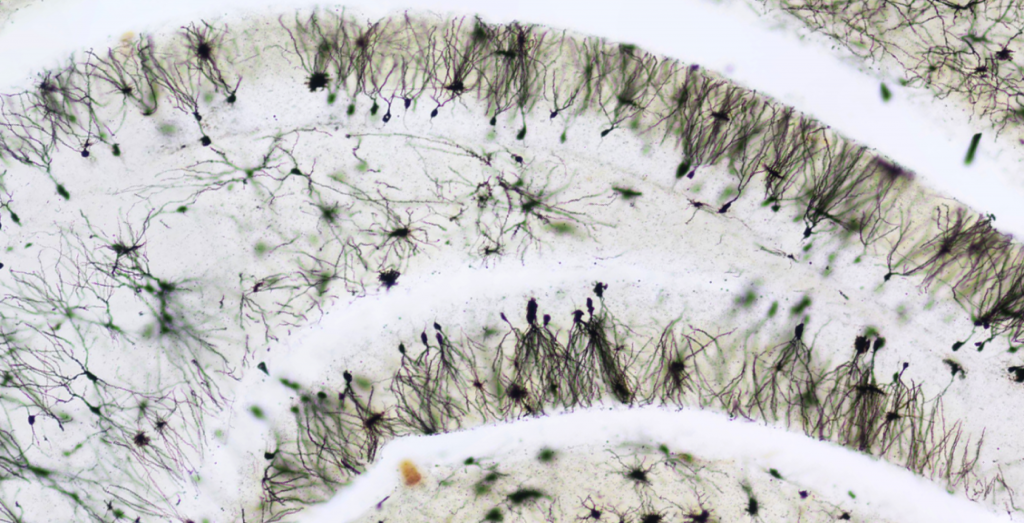
Figure 2. Golgi-stained dentate granule neurons in the dentate gyrus of the Ts65Dn mouse model of Down syndrome. These neurons play a significant role in contextual learning in mice and undergo significant degeneration in the Ts65Dn mouse model of Down syndrome.
Neurolucida was also used in the beginning of the study to compare dendritic length and branching of dentate granule cells in wild type mice (a typical mouse found in nature) and in the Down syndrome mouse model. The researchers found that the average dendritic length is shorter in Ts65Dn mice comapared to controls, and then showed this result with Fan-In Diagrams generated with Neurolucida.

Figure 3. Fan-In Diagram of dendrites of dentate granule cells in the molecular layer of the dentate gyrus visualized using Neurolucida. It depicts the branching pattern of dentate granule cells in an untreated Down syndrome mouse model. A Fan-In Diagram turns a 3D-model of a neuron into a 2D-model. Data are collected in x,y, and z directions and the Fan-In Diagram enables at user to place all neurons along their long axis at the center a 2D plane for comparison.
The results of this paper suggest that focusing on β2ARs is an effective strategy for improving cognitive function in Down syndrome mice. Since it’s already widely used in humans, formoterol or similar β2AR agonists, could be candidates for clinical trials to improve cognitive function in people with Down syndrome. Furthermore, it shows that quantifying the extent of dendritic arborization and complexity might be as accurate as measuring the extent of synapses and cell size in biological settings.
V. Dang, B. Medina, D. Das, S. Moghadam, K.J. Martin, B. Lin, P. Naik, D. Patel, R. Nosheny, J. Wesson, A. Salehi. “Formoterol, a Long-Acting β2 Adrenergic Agonist, Improves Cognitive Function and Promotes Dendritic Complexity in a Mouse Model of Down Syndrome” Biological Psychiatry, Published online 03 July 2013.