
Curcumin Lowers Neuroinflammation in Mouse Model
Scientists at Western Sydney University used Stereo Investigator and Neurolucida 360 to quantify cells in a mouse model of neuroinflammation after feeding mice two different curcumin formulations.
Some inflammation is normal in a healthy mammalian brain. But as the brain ages, processes can break down, leading to chronic neuroinflammation. This can develop into Alzheimer’s disease, dementia, and other neurodegenerative diseases.
Scientists at Prof. Gerald Muench’s lab, at Western Sydney University say that curcumin, a substance in the spice turmeric, has the potential to lower inflammation in the brain.
In two recent studies, the researchers, led by Dr. Erika Gyengesi, used Stereo Investigator and Neurolucida 360 to reconstruct and quantify glial cells in the brains of mice after feeding them two different curcumin formulations.
“MBF Bioscience’s software helped us immensely to differentiate and follow the changes caused by chronic microglia activation in various areas of the brain during aging, but also to quantify the effects of different modified curcumin products, which otherwise would have been impossible,” said Dr. Gyengesi.
In a study published February, 2020 in Scientific Reports: “Effects of a solid lipid curcumin particle formulation on chronic activation of microglia and astroglia in the GFAP-IL6 mouse model,” (Ullah et al, 2020), the researchers describe positive results after feeding GFAP-IL6 mice — a mouse model of chronic neuroinflammation — 500 ppm of Longvida®Optimised Curcumin (LC) over a course of six months.
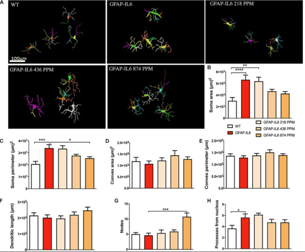
Effect of MC on the morphological characteristics of microglial cells in the hippocampus. (A) Morphological assessment of reactive and non-reactive microglia in the hippocampus. (B–H) Microglia in the inflamed mice have significantly larger soma area, soma perimeter and processes compared with the WT mice. High dose MC significantly reduced soma area and soma perimeter compared with GFAP-IL6 mice. However, the same high dose MC significantly increased the number of nodes compared with the GFAP-Il6 mice. It has no effect on the convex area, convex perimeter, dendritic length and number of processes. Significance = *p < 0.05, **p < 0.001, ***p < 0.0001, ****p < 0.0001.
Stereological analysis of the mouse brains revealed lower levels of activated microglia in the hippocampus (26 percent less) and in the cerebellum (48 percent less) in GFAP-IL6 mice that were fed the curcumin diet, compared to GFAP-IL6 mice fed a normal diet. They also quantified astrocytes — another cell type activated in response to neuroinflammation, finding decreased levels in the hippocampus (30 percent less). TSPO+ cells — another marker of brain inflammation, decreased as well (by 24 percent in the hippocampus and 31 percent in the cerebellum) in the experimental mice compared to controls.
Dr. Gyengesi and her team then checked to see what effect the curcumin formulation had on cell morphology. Using Neurolucida 360 they reconstructed 16 to 20 astrocytes in the hippocampus of each brain of the four different cohorts (wild type normal-fed, wild type LC-fed, GFAP-IL6 normal-fed, GFAP-IL6 LC fed).
They found that in GFAP-IL6 mice, LC decreased “dendritic length of microglia and the convex area, convex perimeter, dendritic length, nodes and number of processes of astrocytes in the hippocampus.” The curcumin formulation also decreased the unusually enlarged soma area and perimeter of neurons in the cerebellum. Increased pre- and postsynaptic protein levels and improved balance were observed as well in LC-fed GFAP-IL6 mice.
In another study, published by the group earlier this year, in the journal Frontiers in Neuroscience, “Evaluation of Phytosomal Curcumin as an Anti-inflammatory Agent for Chronic Glial Activation in the GFAP-IL6 Mouse Model” (Ullah et al, 2020), the researchers tested a different curcumin formulation. This time, they fed GFAP-IL6 mice a soy-lecithin based phytosomal curcumin formulation (Meriva®curcumin).
After feeding the GFAP-IL6 mice three doses of Meriva curcumin over a period of just four weeks, they saw promising results. They quantified lower numbers of activated microglia in the hippocampus (26.2 percent less) and in the cerebellum (48 percent less) compared to those in the population of GFAP-IL6 mice, which was fed a normal diet. Lower levels of GFAP+ astrocytes were also observed in this group.
As in the previous study, the scientists witnessed morphological differences in the mice fed the curcumin diet, including a decrease in the size of the already enlarged soma.
“Using Neurolucida and Stereo Investigator, we have demonstrated that various curcumin formulations with increased bioavailablity have the capability to attenuate chronic inflammatory pathology, by not only reducing activated glial numbers but also reversing their activated morphological state,” said Dr. Gyengesi.
Citations:
Ullah F, Asgarov R, Venigalla M, Liang H, Niedermayer G, Münch G, Gyengesi E. Effects of a solid lipid curcumin particle formulation on chronic activation of microglia and astroglia in the GFAP-IL6 mouse model. Sci Rep. 2020;10(1):2365. Published 2020 Feb 11. doi:10.1038/s41598-020-58838-2 https://pubmed.ncbi.nlm.nih.gov/32047191/
Ullah F, Liang H, Niedermayer G, Münch G, Gyengesi E. Evaluation of Phytosomal Curcumin as an Anti-inflammatory Agent for Chronic Glial Activation in the GFAP-IL6 Mouse Model. Front Neurosci. 2020;14:170. Published 2020 Mar 12. doi:10.3389/fnins.2020.00170 https://pubmed.ncbi.nlm.nih.gov/32226360/