
A complete guide to imaging and analyzing spines and neurons with Neurolucida 360
Following a well-designed protocol is essential to achieving accurate and consistent results in scientific research. Now, scientists using Neurolucida 360 for dendritic spine and neuron analysis can follow a published set of guidelines to ensure optimal confocal data series for proper dendritic spine quantification and neuron reconstruction. The paper, written by MBF Bioscience scientists and researchers from the Icahn School of Medicine at Mount Sinai in New York, was published in Current Protocols in Neuroscience.
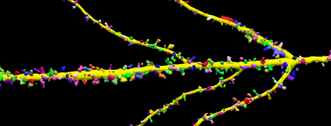
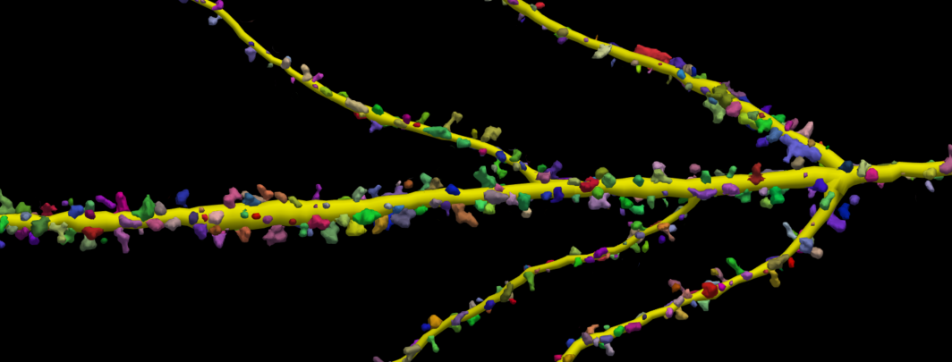
The four protocols describe best practices for imaging and analyzing dendritic spines and entire neurons. Clearly laid out procedures specify necessary materials, image acquisition techniques, and analysis procedures with Neurolucida 360.
Imaging technique is crucial to obtaining unbiased, reproducible results. Clear, crisp images captured with an appropriate z-interval will make analysis with Neurolucida 360 easier and more accurate. Throughout the paper, the authors emphasize the importance of image scaling parameters and unbiased sampling for achieving repeatable results. They also discuss the benefits of correcting optical distortion, especially in the Z-plane, with deconvolution to acquire clear images – a process critical to getting the most accurate representation of dendrites and spines.
Dendritic spine analysis is traditionally performed through tedious, time-consuming manual techniques. According to the paper, this has spawned a growing interest in a more efficient solution for spine quantification and morphological analysis like the one Neurolucida 360 provides. A software platform for automatic neuron reconstruction and spine detection in a 3D environment, Neurolucida 360 offers a variety of benefits, including:
- Fast and accurate spine detection and neuron reconstruction
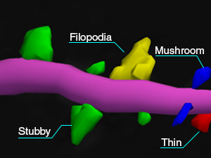
- Accurate spine classification and length quantification using a five-point segment that more accurately models the spine backbone.
- 3 user-guided and automatic algorithms to accurately model neurons visualized with multiple methodologies and imaging techniques.
- A large number of metrics, including volume, length, and surface area.
“We believe that the new quantitative software package, Neurolucida 360, provides the neuroscience research community with the ability to perform higher throughput automated 3D quantitative light microscopy spine analysis under standardized conditions to accelerate the characterization of dendritic spines with greater objectivity and reliability,” (Dickstein, et al. 2016)
The full paper can be found here.
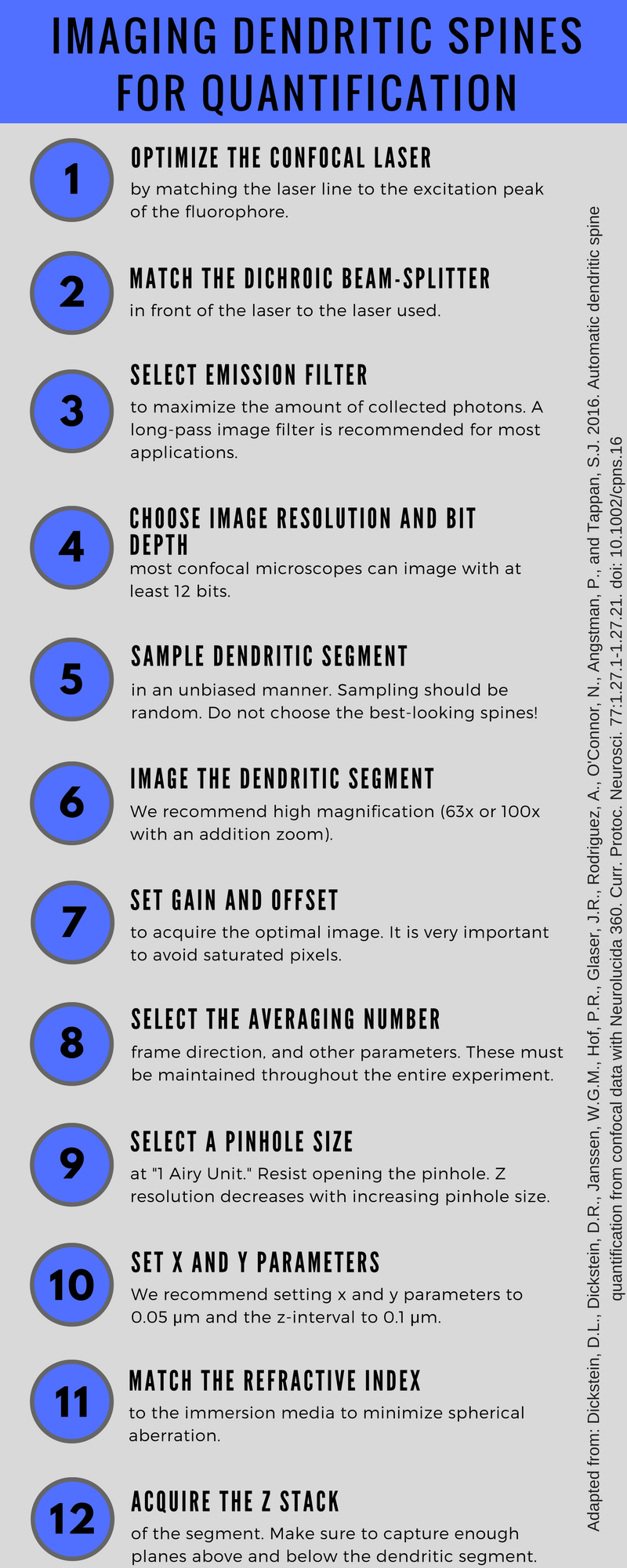
An infographic quickly outlines Protocol 1: Imaging of fluorescently labeled dendritic segments. Use this as a quick reference tool in your lab (right-click on it to save as an image):
Dickstein, D.L., Dickstein, D.R., Janssen, W.G.M., Hof, P.R., Glaser, J.R., Rodriguez, A., O’Connor, N., Angstman, P., and Tappan, S.J. 2016. Automatic dendritic spine quantification from confocal data with Neurolucida 360. Curr. Protoc. Neurosci. 77:1.27.1-1.27.21. doi: 10.1002/cpns.16