
Dying neurons in Alzheimer’s patients show signs of improvement after gene therapy
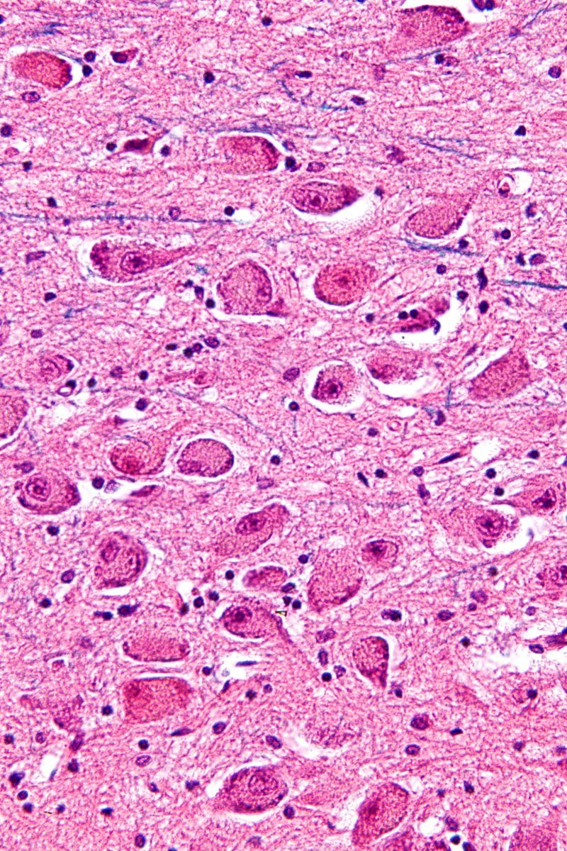
Micrograph of cholinergic neurons in the nucleus basalis of Meynert. Image from Wikipedia.
Cholinergic neurons degenerate at devastating rates in Alzheimer’s disease, but Dr. Mark Tuszynski and his team at the University of California, San Diego may have found a way to slow the decline.
Their study, published in JAMA Neurology, reports that nerve growth factor gene therapy increased the size, axonal sprouting, and signaling of cholinergic neurons in 10 Alzheimer’s disease patients.
The patients were enrolled in a clinical trial between 2001 and 2012. Ex vivo and in vivo methods of gene therapy were used to deliver nerve growth factor – a protein that protects neurons and stimulates growth – to the patients. Eight received an implant of their own skin cells that were genetically modified to express nerve growth factor (ex vivo ) and two patients received injections that induced neurons already in the brain to express nerve growth factor (in vivo). In all 10 patients, gene therapy was delivered to the nucleus basalis of Meynert – part of the basal forebrain rich in cholinergic neurons that undergoes degeneration during Alzheimer’s disease.
The patients’ survival time ranged from one to 10 years. After they had died, researchers analyzed the effects of nerve growth factor on cholinergic neurons.
The axons of cholinergic neurons, labeled with p75, grew toward the source of the nerve growth factor in all 10 patients. To determine if there was a change in cell size, researchers used the nucleator probe in Stereo Investigator to analyze cholinergic neurons of 3 patients who received gene therapy via the ex vivo method in one hemisphere – the other hemisphere was used as a control. Results from Stereo Investigator showed that cell bodies were larger in the treated hemisphere vs. the untreated hemisphere.
Finally, to find out if nerve growth factor induced signaling within cells, the researchers analyzed the amount of CREB and c-fos – markers for cell activation – in 2 patients who received nerve growth factor in vivo. An elevated amount of CREB and c-fos was found when compared to control regions. Neurons exhibiting tau pathology also expressed nerve growth factor, indicating that degenerating cells could respond to nerve growth factor gene therapy.
A phase 2 clinical study is currently under way to report cognitive outcomes in patients with Alzheimer’s disease.
“Collectively, these anatomical findings support the rationale for clinical trials to test the hypothesis that sustained growth factor delivery over time can reduce cell degeneration and stimulate cell function in chronic neurodegenerative disorders, thereby slowing functional decline,” Tuszynski, et al.
Tuszynski, M.H., Yang, J.H., Pay, M.M., Masliah, E., Barba, D., U, H.S., Conner, J.M., Kobalka, P., Roy, S., and Nagahara A.H. (2015). Nerve Growth Factor Gene Therapy: Activation of Neuronal Responses in Alzheimer Disease. JAMA Neurology, published online August 24, 2015. DOI: 10.1001/jamaneurol.2015.1807.