New Zealand Scientists Use Stereo Investigator to Develop a New Model for Human Extreme Prematurity
Each year, nearly ninety thousand children are born extremely premature in the United States – that is, before 28 weeks gestation. Most of them survive, but about half the survivors suffer from severe health problems throughout their childhood and into adulthood, including learning and behavioral disorders such as ADHD.
“Treatment options are clearly urgently required to prevent the brain damage and associated memory deficits that follow extremely premature birth,” say the authors of a study published last month in the Journal of Neuroscience.
Treatment options are limited, the authors say, because current small animal models fall short in their mimicry of the extremely premature human brain. However, the researchers from the University of Otago in New Zealand have come up with a new animal model for human extreme prematurity, which they say more closely resembles the pathological and behavioral deficits seen among this populationThe research team developed the model by exposing rats to an oxygen deficient environment at one to three days of age, a period equivalent to 24-26 weeks human gestation.
By performing behavioral assessments and a stereological analysis of several different regions of the rat brains, the scientists saw similarities in “short and long-term white matter neuropathological injury, gray matter volume loss, and long-term memory deficits” between the rats and children born extremely prematurely.

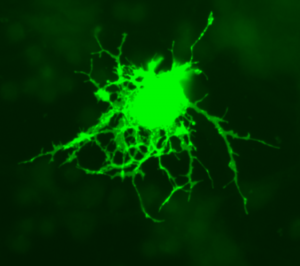
Oligodendrocytes, pictured here with a green fluorescent protein, form a myelin sheath – the insulation around axons. The extremely premature brain features a lower number of pre-oligodendrocytes, thereby decreasing myelination, a characteristic which has been associated with ADHD. Image courtesy of Wikimedia Commons.
They used the Cavalieri, optical fractionator, optical disector, and nucleator probes in Stereo Investigator to quantify different elements of brain tissue. The researchers found that, compared to controls, the hypoxic rats had a lower number of pre-oligodendrocytes at four-days-old, and at 14-days-old displayed less cerebral white matter volume and myelin, as well as less cerebral cortical and striatal gray matter volume without neuronal loss – pathological characteristics also present in the brains of humans born extremely premature.
Behavioral tests showed the mice mimicked behavioral characteristics in children and adults born extremely prematurely, displaying memory deficits and ADHD-like hyperactivity.
“This new rat model provides a clinically relevant tool to investigate numerous cellular, molecular, and therapeutic questions on brain injury attributable to extreme prematurity,” the authors say.
Oorschot, Dorothy E., Voss, Logan, Covey, Matthew V., Goddard, Liping, Huang, William, Birchall, Penny, Bilkey, David K. and Kohe, Sarah E. (2013). Spectrum of Short- and Long-Term Brain Pathology and Long-Term Behavioral Deficits in Male Repeated Hypoxic Rats Closely Resembling Human Extreme Prematurity. The Journal of Neuroscience, 33(29), 11863–11877. doi: 10.1523/jneurosci.0342-12.2013.
Image courtesy: Wikimedia Commons


