In Focus: Dr. Clara Thore
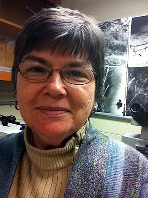
Who: Clara R. Thore, Ph.D., Computer Imaging Specialist
Where she works: The Microvascular Research Lab, Department of Diagnostic Radiology, Wake Forest University School of Medicine , Winston-Salem, North Carolina
Research focus: Microvascular changes in the brain in diseases of aging, such as vascular dementia, leukoaraiosis, and Alzheimer’s disease.
MBF Bioscience software used: Stereo Investigator
Research methods at a glance: Dr. Thore and her fellow researchers have come a long way from their early studies involving scanning AP stained sections on a flatbed scanner to measure vessel density. Today, they use stereological methods, which provide more accurate data and take less time.
What she does: Dr. Thore studies the brain’s tiniest blood vessels. She examines human autopsy material and highly sensitive archival tissues for her research on Alzheimer’s disease, vascular dementia, and other diseases of aging.
At her lab, The Brain Microvascular Pathology Laboratory, she and her colleagues use Stereo Investigator to quantitate vascular densities in the human brain and the brains of experimental animals. One hundred-micrometer-thick sections of celloidin-embedded tissue blocks with arteries, arterioles, and capillaries marked with AP staining provide Dr. Thore with a 3D view of her subject. By implementing stereology, immunohistochemistry, and confocal microscopy techniques, she studies changes to the cerebral microvascular system.
Previously, Dr. Thore and her team measured vessel density by scanning the AP stained sections on a flatbed scanner, which she said provided crude area fraction data. They then captured digital images from sections and used algorithms to process the images to binary in order to quantitate vessel density as area fraction. “The image processing introduced variance, was time consuming, and did not account for the depth of the captured field,” she explained. Dr. Thore and her colleagues began using stereological methods, which they found avoided these problems by providing random systematic sampling.
“The installation of the Stereo Investigator system by MBF Biosciences has made a huge difference in our quantitative approach,” said Dr. Thore. “By using the built-in Stereo Investigator Space Balls probe, we no longer have to be concerned with the directionality of vessels in our sections. Stereological quantization is done real-time, which has decreased the time needed per specimen and allowed us to increase the number of areas and slides sampled.”
“In Focus” is a new series that spotlights scientists who use MBF Bioscience products in their work. Thank you Dr. Clara Thore for participating.
For the latest news about MBF Bioscience and our customers, fan us on Facebook and follow us on Twitter