In Memoriam: Edmund M. Glaser, PhD
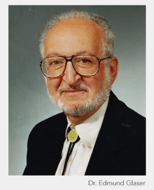
Dr. Edmund Glaser devoted his career of more than four decades to the field of neuroscience. Most notably, in 1963, he co-invented computer microscopy, a pioneering method of quantifying the brain’s morphometry. This technology, for the first time, applied computer techniques to the neuroanatomical world, permitting scientists to precisely quantify the brain’s three-dimensional structure. It simplified time-consuming, inexact classical methodologies in an efficient and cost-effective method. By 1995, the year of Dr. Glaser’s retirement, computer microscopy had been adopted by thousands of neuroscience laboratories throughout the world.
 Dr. Glaser started his college education in his hometown, New York City, studying Electrical Engineering at The Cooper Union. In the midst of his college career, he was drafted and served in the U.S. Army during WWII. His duty took him to Nuremberg, where he was a sound recordist and photographer who documented the Medical War Trials of infamous Nazi physicians. After his military service, he completed his bachelor’s degree in 1949. After doing early project work in communications systems and guided missiles for the U.S. Air Force, he soon became attracted to the emerging fields of computing, information theory, and artificial intelligence. In 1952, he returned to his academic studies in engineering. He received a PhD from Johns Hopkins University in 1960 and then secured a postdoctoral fellowship in its Department of Physiology in the School of Medicine.
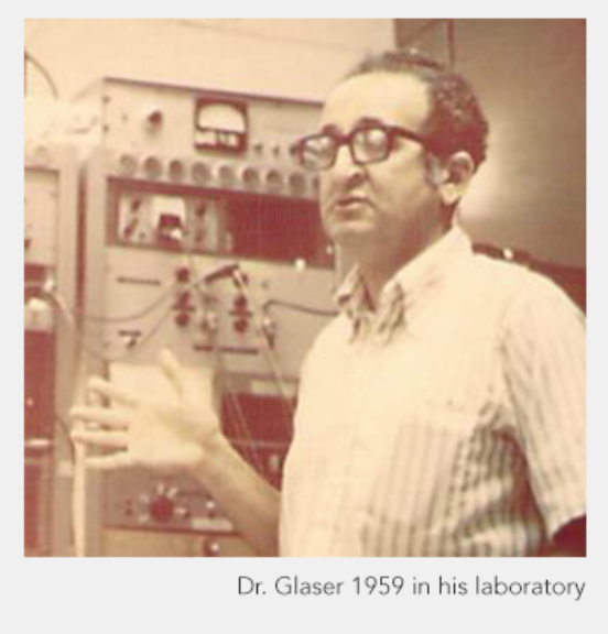
In 1963, Dr. Glaser teamed up with Dr. Hendrik Van der Loos, a neuroanatomist at Johns Hopkins, to study the complex morphology of the brain’s cerebral cortex. They encountered the shortcomings of the time’s tedious neuroanatomical techniques and noted the need to revamp the prevailing methods of analyzing neuron morphology and neuronal networks within the cerebral cortex. It was then that they formulated the design and the construction of the first computer microscope.
Computer microscopy at that time was based on the use of analog computer technology. Glaser and Van der Loos demonstrated the great improvements that could made in neuroanatomy by adapting computer technology, it showed the practical way to represent the brain’s structure in its intrinsic three-dimensional reality. In so doing, the quantification of neuroanatomy was wholly revolutionized. Tracing neuronal structures was reduced from hours to minutes and measurement precision was able to achieve fractional micron accuracy. What is more, large assemblies of neuronal networks could be examined in quantitative detail in three dimensions.
The fundamental discovery by Glaser and Van der Loos of the so-called autapse, a synapse between the axon and a dendrite of the same neuron, was made possible by their new computer microscope technology. The autapse could not have been discovered using classical viewing techniques, as the observer lacked the ability to relate the connections between separate parts of a neuron at the same time. To this day, the autapse remains a poorly understood part of neuronal morphology.
 When the full description of their original computer microscope was published in 1965 in the IEEE Transactions on Biomedical Engineering, Robert Schoenfeld, with great foresight, wrote in an accompanying editorial, “Some observers believe that we are now entering upon a Great Age of Biology, and that the remainder of this century will be … spent doing for biology … what the previous age has done for the physical sciences. … A foreshadowing of the development of computer techniques in biology appears in a paper by E. M. Glaser and H. Van der Loos. It describes a semi‐automatic computer‐microscope that can be used in quantitative neuroanatomical studies. … This new instrument … integrates sensing transducers and an analog computer with the mechanical stage of a light microscope for measuring micro distances.”
Glaser and Van der Loos coined the term “computer microscopy” at a time when analog computers were still popular, and digital computers were a rarity—monster‐like in size and snail‐like in speed. The further development of computer microscopy and its gradual adoption by anatomists took place over an interval of more than 20 years. Digital technology was eventually adopted, and Glaser went on to invent the Image-Combining Computer Microscope, which combined all the aspects of computer microscopy in a single instrument, and he subsequently patented it.
In 1987, Dr. Glaser co-founded the company MicroBrightField (now MBF Bioscience) with his son, Jacob (Jack) Glaser. Their commercial microscope system, named Neurolucida, became known for its ability to significantly enhance neuroanatomical research. Ultimately, this technology would be also applied to stereological analysis, in the Stereo Investigator system, to help quantify the properties of brain morphology. In the ensuing years, these systems have supported numerous key findings in neuroscience, spanning more than 1,000 laboratories across the world in more than 15,000 published papers.


