
Celebrating 50 years of Computer Microscopy (1963 – 2013) Part II: The Impact of the Computer Microscope on Neuroanatomical Analysis
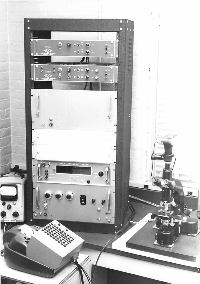
Overall view of the computer microscope developed by Drs. van der Loos and Glaser (circa 1965).
Prior to the computer microscope era, quantitative neuro-anatomical studies were performed using the camera lucida method, an optical method allowing the scientist to see the neurons as if reflected on the piece of paper on which she will trace. These studies were painstaking and extremely time-consuming. Van der Loos and Glaser’s computer microscope was truly groundbreaking in that it reduced analysis times dramatically, from twenty-four hours with the camera lucida and hand calculation techniques, to only thirty minutes with the computer microscope. Accuracy was also greatly improved thanks to accurate distance measurements in all three coordinate axes.
Interestingly, the development of the computer microscope coincided with the important discovery of the Green Fluorescent Protein (GFP) that would enable the study of neurons in vivo for the first time, and pave the way for the discovery and engineering of more fluorescent markers. Numerous studies involve the use of GFP today, including a study by French scientists who labeled adult-born neurons in the dentate gyrus of the hippocampus with a GFP retrovirus to study their plasticity and performed morphometric analyses with Neurolucida.
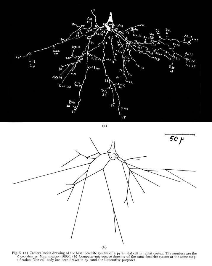
From “A Semi-Automatic Computer-Microscope for the Analysis of Neuronal Morphology,” by E. M. Glaser and H. van der Loos, 1965, Biomedical Engineering, IEEE Transactions 1, p. 26.
Since Drs. Glaser and van der Loos invented the computer microscope, there have been many advances in microscope and computer technology and Neurolucida has grown along with them. The computer microscope is still the preferred way to trace and analyze neurons, fifty years after its invention. For more information on the original computer microscope for neuron tracing, read Drs. Glaser and van der Loos’ paper “A Semi-Automatic Computer-Microscope for the Analysis of Neuronal Morphology” published in 1965. This paper was singled out by IEEE editors as a great advance for what they call the Great Age of Biology.
But was the value of the computer microscope, besides a few accolades, recognized by the scientific community in the sixties? According to Edmund Glaser, “little attention was paid to it for the next 10 years. Nonetheless, we kept on working on it as did some other neuroanatomists. Our own additional contributions and those of some others pushed it into the digital world.” We’ll explore some of the contributions made in the seventies in our next blog post.