
Vesselucida Helps Researchers Quantify Post-Injury Capillary Damage and Regeneration
Our health depends on the ability of blood vessels to deliver nutrients and remove metabolic byproducts from organs and muscle systems. But what happens to this delicately balanced process after traumatic injury? Scientists generally understand that skeletal muscles can regenerate, but little is known about how this happens at the level of our microvasculature.
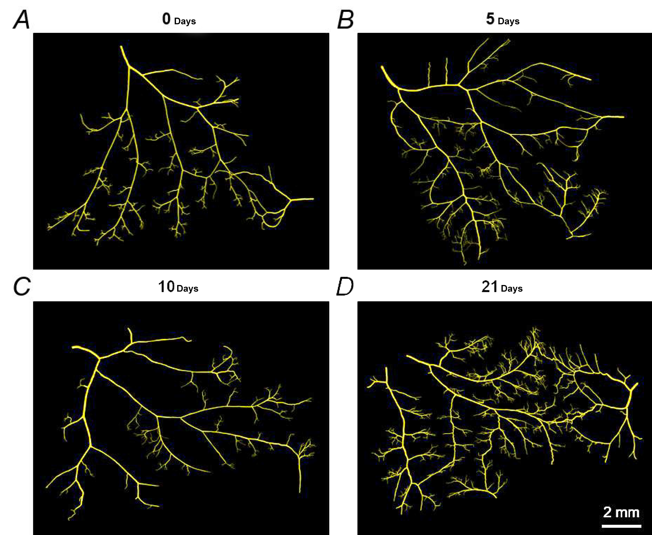
Representative maps of resistance networks from feed artery to terminal
In a study published in the Journal of Physiology, researchers at the University of Missouri, Columbia, describe short- and long-term capillary damage and recovery after acute skeletal muscle injury. The researchers observed that two to three days after injury, surviving microvessel fragments began to sprout new capillaries, and that five days post-injury new functional capillary networks formed.
In this study, researchers used Vesselucida from MBF Bioscience to characterize changes in skeletal microvasculature throughout a mouse model of muscle injury. 3D reconstructions of injured and recovering vessels in whole-mount preparations of the gluteus maximus muscle at various time points reveal the chronological degeneration and remodeling of the vascular network before and after injury.
“Using Vesselucida, we were able to assess changes in resistance network architecture during muscle regeneration for the first time,” said Dr. Nicole Jacobsen. “We were limited by other imaging methods due to the network size and location of arteriolar networks within skeletal muscle. Vesselucida uniquely enabled us to reconstruct and analyze intact arteriolar networks in 3 dimensions with micrometer resolution over distances spanning millimeters to centimeters.”
When quantifying segments and overall length of capillaries in Vesselucida, Dr. Jacobsen and her team binned the data based on vessel diameter to consider changes in vasculature of varying sizes. This revealed injury and recovery-related morphological changes in capillaries (five to ten micrometers in diameter), but not in arterioles and venules.
This study is unique in demonstrating significant microvasculature damage and repair in a model of acute injury using thick tissue sections. In previous studies, thin sections have been used selectively to show cross-sections of capillaries with a measurement bias based on their proximity to muscle-cell intersects (Jacobsen, et. al. 2021).
This work demonstrates the unique power of using Vesselucida to trace capillaries and other vessels in thick sections, while avoiding the bias inherent in thin section measurements, to more accurately characterize vascular networks.
Jacobsen NL, Norton CE, Shaw RL, Cornelison D, Segal SS. Myofibre injury induces capillary disruption and regeneration of disorganized microvascular networks. J Physiol. 2021 Nov 11. doi: 10.1113/JP282292.