Cincinnati Scientists Use Neurolucida in Epilepsy Study
Scientists hypothesize that seizures occur because brain cells fire in places they’re not supposed to. Dentate granule cells (DGCs), a type of neuron born throughout adulthood, sometimes migrate into a different region of the dentate gyrus, a part of the hippocampus. These abnormal newborn cells sprout axons called “mossy fibers” that form connections with neighboring DGCs in the inner molecular layer, causing synaptic changes that wouldn’t normally occur in healthy brains.
Much research has been done on this phenomenon, but neuroscientists still struggle to understand what exactly its relationship is with epilepsy.
A new study by researchers at the Cincinnati Children’s Hospital Medical Center validates hypotheses about the role of abnormal DGCs in epilepsy. In their study of a transgenic mouse model of temporal lobe epilepsy (TLE), the scientists observed a relationship between the presence of deviant DGCs and seizure frequency.
“Animals possessing a substantial percentage of ectopic newborn DGCs and robust [mossy fiber sprouting] exhibited seizures more often than epileptic animals in which a smaller percentage of newborn cells developed abnormally,” they say in their paper, published last month in the Journal of Neuroscience.
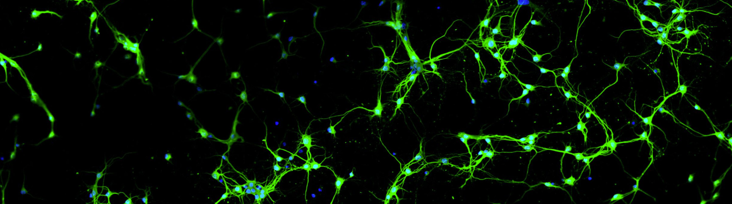
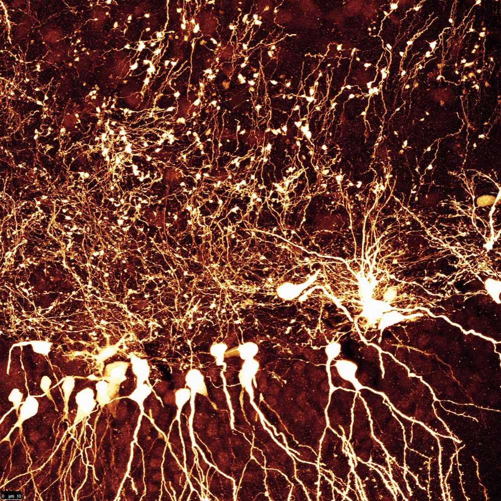
The research team quantified newborn DGCs by generating computer images of both the dorsal and ventral hippocampi of eight mice. They created three-dimensional confocal z-stacks with images of cells expressing the GFP gene, a marker for neurogenesis. By loading these image stacks into Neurolucida, they were able to count the number of newborn DGCs with basal dendrites projecting into the dentate gyrus’ inner layer, the dentate hilus, where prior research has shown they become targets for mossy fiber innervation. The scientists found mice experienced longer seizures when they had more of these kinds of cells.
“I’m a huge fan of Neurolucida’s cell tracing and auto-detection features, which greatly assisted me in characterizing the morphology of abnormal neurons,” first author Michael Hester said. “Neurolucida is easily learned, intuitive, and was clearly designed by scientists.”
They also used Neurolucida to count mossy cells in the dentate hilus, determining that animals with low numbers of hilar mossy cells experienced the highest rate of seizures, while mice with the fewest seizures “contained a dense network of mossy cells in the hilar area.”
“These findings are important because they suggest that – in addition to regulating the incidence of seizures – dentate pathology might also regulate seizure severity,” the authors say. “Together, the present findings provide new correlative evidence suggesting that aberrant neuronal integration potentially acting in concert with cell loss are key steps in temporal lobe epileptogenesis.”
Hester, M.S., Danzer, S.C., (2013). Accumulation of Abnormal Adult-Generated Hippocampal Granule Cells Predicts Seizure Frequency and Severity. The Journal of Neuroscience, 33(21): 8926-8936; doi: 10.1523/ JNEUROSCI.5161-12.2013
[Images of Adult-born dentate granule cells and abnormal glial cells in epileptic mice provided by the study’s authors.]