NeuroDeblur®
Deconvolution Software for 3D Microscopy - Groundbreaking Speed and Quality - at a Reasonable Price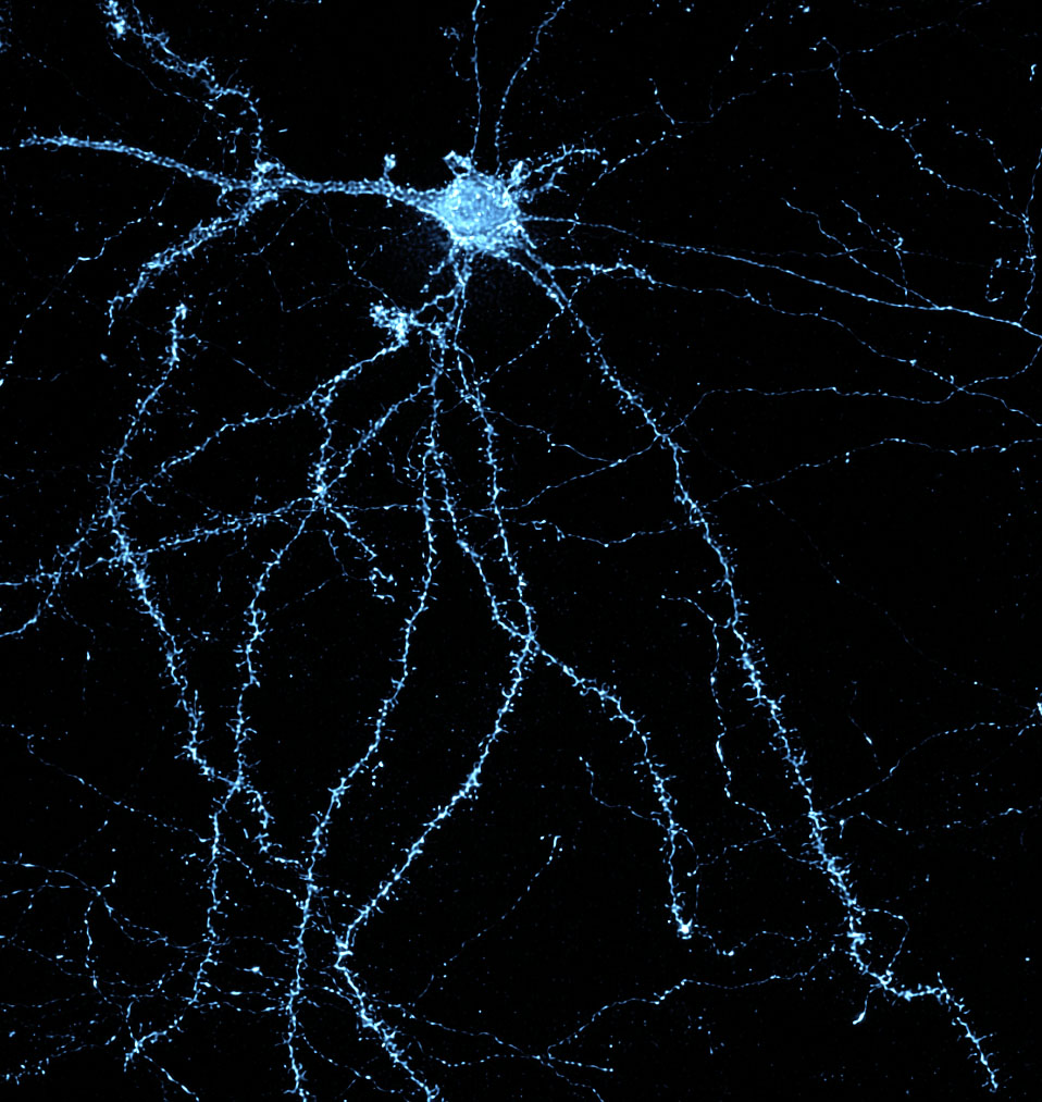
NeuroDeblur is the groundbreaking solution for deconvolution and artifact removal in microscopy images, particularly for large data sets from light sheet and confocal microscopes. Engineered by experts in microscopy and deconvolution, NeuroDeblur produces optimal results from a wide array of microscopes, further enhancing images from even the best microscopes.
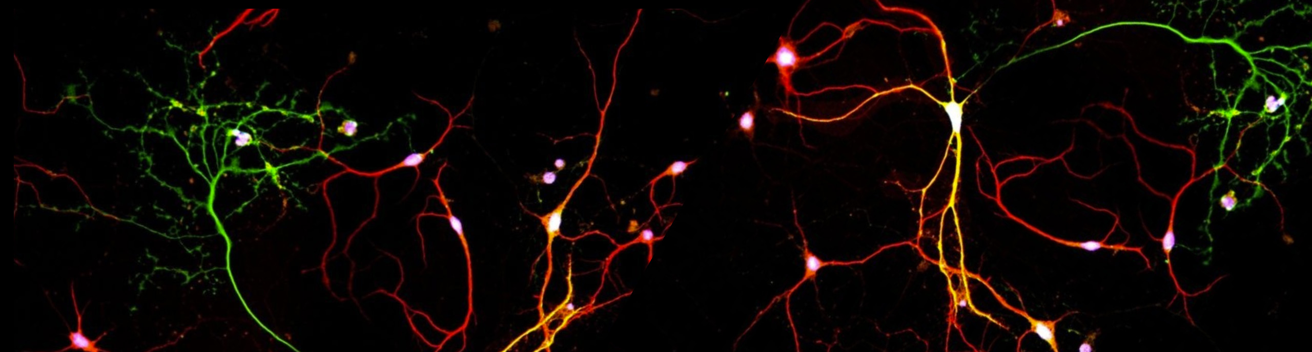
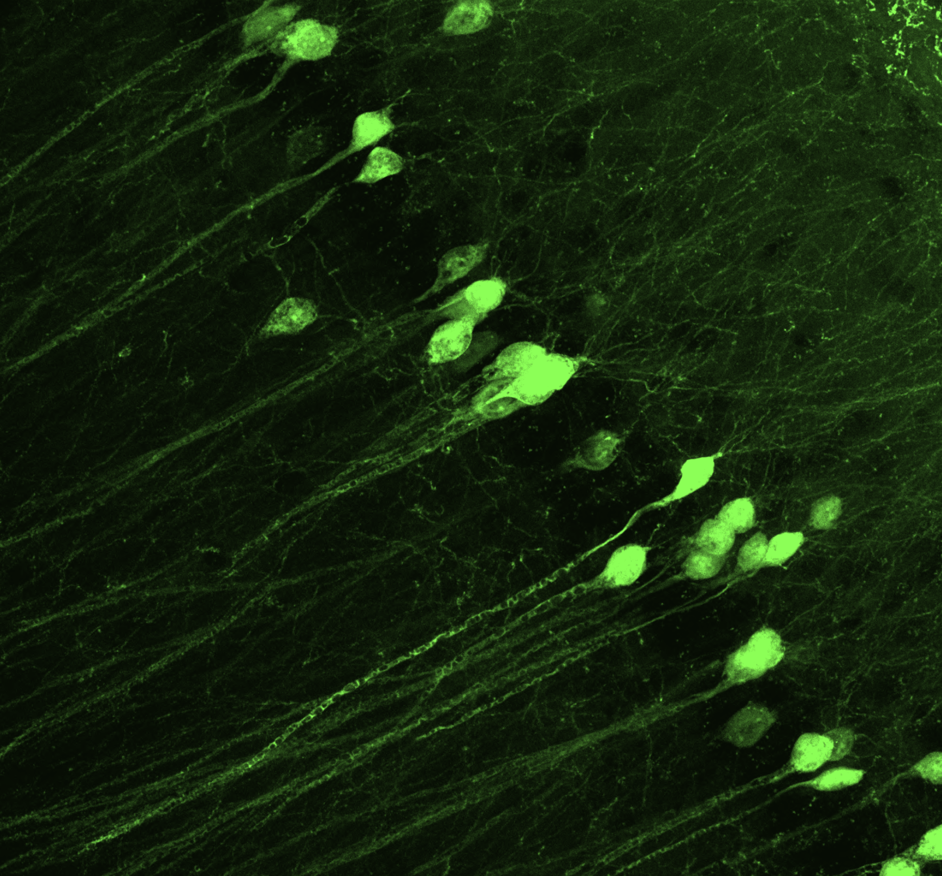
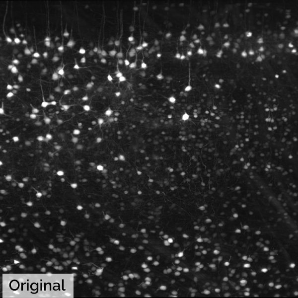
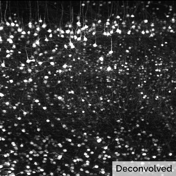
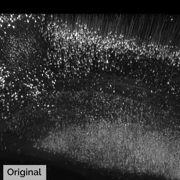
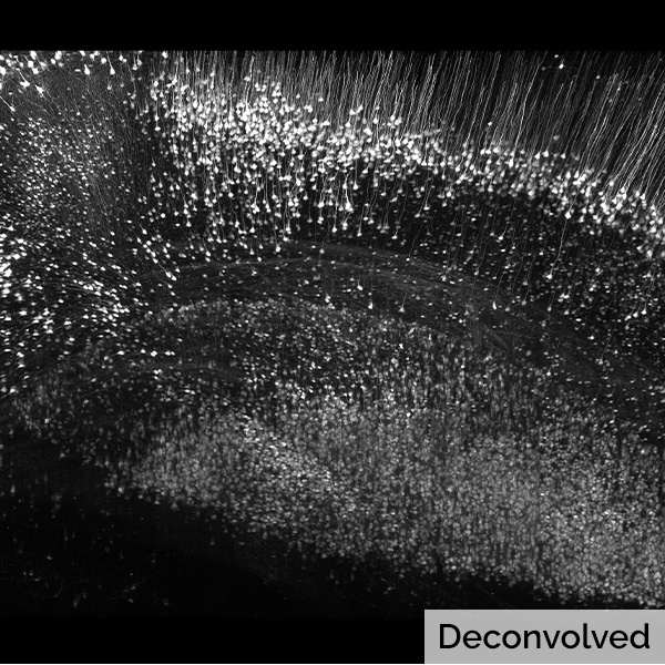
NeuroDeblur significantly enhances the visibility of intricate details of cells such as neurons, glia and subcellular structures, such as axons and dendritic spines. The software offers non-blind deconvolution with a synthetic point spread function (PSF) optimized for each imaging mode, as well as the option to use a measured PSF. NeuroDeblur was originally developed for processing extremely large image stacks obtained from light-sheet microscopy. However, its advanced block-wise computational processing capabilities enable it to handle datasets of virtually unlimited size. Unwanted image background is removed using an advanced 3D rolling ball filtering algorithm. By leveraging parallelization and optional GPU acceleration on NVIDIA graphics cards, NeuroDeblur achieves remarkable processing speeds. For example, on a PC equipped with a mid-range gaming graphics card, a 3D stack containing approximately 1 billion voxels can be deconvolved within 5-10 minutes. This powerful combination of advanced features and efficient performance makes NeuroDeblur an indispensable tool for researchers seeking to unravel the intricacies of microscopic structures with unprecedented clarity and speed.
Key Features:
- Statistically rigorous image restoration at an affordable price, supporting popular imaging modalities like light sheet, confocal, spinning disk, and widefield microscopy.
- GPU-accelerated deconvolution that produces clear images in minutes rather than hours, compatible with most NVIDIA graphics cards and capable of processing even the largest light sheet data sets.
- Excellent deconvolution results for light-sheet and confocal microscopy without the need to measure the imaging system's point spread function (PSF) along with sophisticated adaptive image background removal and stripe-artifact removal for light-sheet microscopy data.
- Supports almost all microscopic image file formats without added cost and works with images from most common microscope manufacturers.
- Easy-to-use GUI with 3D visualization and a powerful command-line interface for embedding deconvolution into complex workflows.
Additional features include deconvolution using measured PSFs, powerful adaptive background artifact correction, fast multiprocessor-aware deconvolution with noise minimization, batch deconvolution for automated imaging workflows, automatic block-wise processing of large data sets on computers with limited RAM, ROI and color channel processing for proprietary 4D or 5D input formats, color channel mixing for optimized display of deconvolved results, and image post-processing by adaptive histogram equalization (CLAHE) and unsharp masking.
- Excellent deconvolution results with light sheet and confocal microscopy without or without PSF measurements
- Deconvolution using matching accurate PSF models for light sheet and confocal microscopy
- Deconvolution using a PSF measured from the imaging system
Powerful adaptive background artifact correction - Fast multiprocessor aware deconvolution with excellent regularizing for noise minimization
Ultra-fast GPU based processing with modern Nvidia graphic cards (> 100 million voxels/min) - Batch deconvolution for building an automated imaging workflow
Automatic block-wise processing of large data sets even on computers with limited RAM - Sophisticated stripe-artifact removal for light sheet microscopy data
Side-by-side comparison of original and deconvolved data in synchronized display windows - Process select ROIs and color channels of proprietary 4D- or 5D-input formats (e.g. Zeiss, Evident, Leica, etc).
- Optimize the display of deconvolved results via additive and subtractive color channel mixing.
- Image post-processing by adaptive histogram equilibration (CLAHE) and unsharp masking.
Download NeuroDeblur product sheet here.
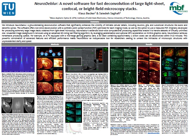
NeuroDeblur: A novel software for fast deconvolution of large light‐sheet, confocal, or bright‐field microscopy stacks


Boeglin, M., E. Leyva-Díaz, et al.
Expression and function of C. elegans UNCP-18, a paralogue of the SM protein UNC-18View Publication

Rentsch, P., T. Egan, et al.
The ratio of M1 to M2 microglia in the striatum determines the severity of L-Dopa-induced dyskinesiasView Publication

Wang, Z., D. Zheng, et al.
Enabling Survival of Transplanted Neural Precursor Cells in the Ischemic BrainView Publication

Villar-Conde, S., V. Astillero-Lopez, et al.
Synaptic involvement of the human amygdala in Parkinson’s diseaseView Publication

Stimpson, C. D., J. B. Smaers, et al.
Evolutionary scaling and cognitive correlates of primate frontal cortex microstructureView Publication

Russ, T., L. Enders, et al.
2,4-Dichlorophenoxyacetic Acid Induces Degeneration of mDA Neurons In VitroView Publication

Olkhova, E. A., C. Bradshaw, et al.
A novel mouse model of mitochondrial disease exhibits juvenile-onset severe neurological impairment due to parvalbumin cell mitochondrial dysfunctionView Publication

Zhang, X., C. Wang, et al.
Analysis of Error Sources in the Lissajous Scanning Trajectory Based on Two-Dimensional MEMS MirrorsView Publication

Lu, J., Behbahani, A.H., Hamburg, L. et al.
"Transforming representations of movement from body- to world-centric space."

"I rarely have encountered a company so committed to support and troubleshooting as MBF."

"MBF Bioscience is extremely responsive to the needs of scientists and is genuinely interested in helping all of us in science do the best job we can."

"I am so happy to be a customer of your company. I always get great help related with your product or not. With the experienced members, you are the best team I've ever met. All of your staff are very kind and helpful. Thank you for your great help and support all the time."

"We’ve been very happy for many years with MBF products and the course of upgrades and improvements. Your service department is outstanding. I have gotten great help from the staff with the software and hardware."

"Our experience with the MBF equipment and especially the MBF people has been outstanding. I cannot speak any higher about their professionalism and attention for our needs."
"MBF provides excellent technical support and helps you to find the best technical tools for your research challenges on morphometry."