Spaceballs
|
|
Length |
You can determine length for many different types of objects (or fibers): tubules, nerve fibers, small blood vessels, surfaces, microvilli, etc. Spaceballs provides a length estimate instead of length-density. Reporting length per region instead of length per volume is more effective because a length-density measure can't account for possible concomitant changes in volume along with length.
|
|
|
|
Thick |
||
|
|
Preferential section |
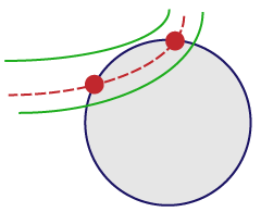
The probe is a sphere or hemisphere virtually embedded in the tissue, and the counted parameter is the number of profiles that transect the edge of the sphere within a defined counting region. Since the surface of a sphere is isotropic, the need for isotropic uniform random (IUR) sections is eliminated.
In Stereo Investigator, Spaceballs is implemented with a fractionator sampling methodology to return an estimate of total length per region. A series of sites are selected within each section by systematic random sampling. At each site, a 3D sphere of constant volume is superimposed upon the slide to represent the intersection sites to be counted. The item to be counted is a "profile," that is, the point where the fiber transects the sphere boundary. If fibers are thick, you must identify the center point of the fiber, and only count locations where the center point transects the sphere outline.
- Thick sections (significantly thicker than the diameter of the tubules/fibers to be measured)
- Structure of interest stained through the depth of the tissue section.
- Unstained tissue transparent enough to see the stained structure throughout the depth of the section.
- Thin focal planes achieved with a high magnification, high numerical aperture lens, generally oil immersion.
- A contour traced around the region of interest.
Use a high power objective lens with a high numerical aperture to focus at the top and bottom of a few sites throughout your sections to get a rough idea of the section thickness. This value can be edited later, but an approximate value is needed for setting up the 3D sampling boxes and guard zones.
You don't need to define a counting frame for the Spaceballs probe. However, the previously defined counting frame size does have an influence on the grid size if the counting frame is larger than the desired grid.
If Preview SRS Layout does not let you specify a large enough number of sampling sites, click Probes>Define Counting Frame to define a smaller counting frame.
- Click Probes>Preview SRS Layout to preview the arrangement of sampling sites within the tissue section.
If there's more than one contour in the current section, an overview of the data file is presented, and the desired contour should be selected.
Once the settings have been selected, the sampling site layout is shown in the tracing window.
- Click anywhere in the tracing window to end the Preview mode.
- Start the Serial Section Manager and define the first section.
- Draw a contour around the region of interest.
- Click Probes>Preview SRS Layout to set the size of the scan grid.
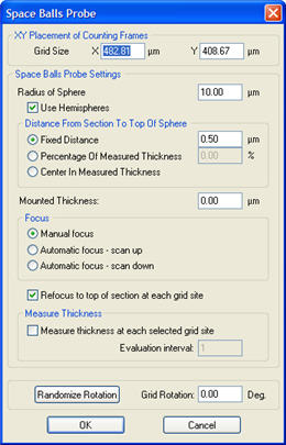
- Click Probes>Spaceballs. The Spaceballs dialog box opens
 . Adjust the settings as needed.
 Settings
If SRS Preview was used, the Scan Grid Size determined in the preview is automatically entered in the XY Placement of Counting Frames field.
Spaceballs Settings: The radius should allow for 2-3 transection events per sampling site. The sphere or hemisphere must be sized to fit entirely within the tissue thickness, including top and bottom guard zones, if they are used.
Select Use Hemispheres to use the top half of a sphere as the sampling probe. Counts that are on the bottom edge of the hemisphere are factored in as 1/2 counts in the results.
Distance from Section Top to Top of Sphere is the top guard zone height. This should be larger than the average thickness of the fibers to be counted. It can also be set to a percentage of the measured section thickness, in which case the program automatically selects measurements of the section thickness at every sampling site.
Mounted Thickness is the actual Z-depth of the sections as mounted on the slides (not cut thickness). Measure it with your microscope before starting. If you are using Stereo Investigator to measure the section thickness at each sampling site, it isn't necessary to enter a value for the mounted section thickness since it is calculated by the program. This value can be edited when viewing results if the initial estimate proves incorrect.
Focus:
- If Refocus to top of section at each grid site is selected, Stereo Investigator prompts you to focus at the top before setting up the counting box at each sampling site.
- If Measure thickness at each selected grid site is selected, the program prompts you to focus at the top of the section and bottom of the section at each sampling site (or at a sample of sites set with Measurement site periodicity).
- Use Measure thickness at each selected grid site for Number Weighted Thickness results.
- Stereo Investigator drives the stage to the first sampling site. The cursor changes to
 , indicating that the plane of focus is above the counting frame.
 - Focus down until a small circle is visible in the center of the field of view. If the grid spacing is small, multiple circles may be visible.
- Select a marker for transections.
- Place a marker at each location where a fiber crosses the currently visible circle.
- If multiple circles are visible, only mark the circle in the center, as the other sites will be systematically visited.
- Focus through the section, marking each intersection between a fiber and the sphere, until all intersections have been counted. The circle will appear larger in each subsequent focal plane to represent the sphere or hemisphere.
- Count as 1/2: This option is available in the right-click menu if Hemispheres are being used and the current focal plane is near the equator of the hemisphere. If a linear feature is at the widest point of the hemisphere, it should be counted as 1/2, since it would also be counted in the lower half of a full sphere.
- Right-click and select Next Scan Site.
- Complete all sampling sites within the section.
- Move the stage to a new section, use the Serial Section Manager to designate a new section, and repeat steps 2–8.
![]() View Overview Layout
View Overview Layout
At any time during the probe run, right-click Preview Spaceballs to see an overview of the probe layout. Stereo Investigator displays the entire file with the sampling boxes overlaid on the tracing. The current sampling box is indicated by flashing. Click anywhere in the Preview window to return to the current sampling site.
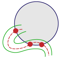
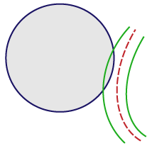
For vessels that appear on the edge of the spaceball, use the “center line” rule:
- If the center line enters and then leaves the probe, mark it twice (at the entrance point and at the exit point).

- If the center line weaves in and out, mark the centerline very time it enters or leaves the spaceball.

- If the center line doesn't enter the spaceball, don’t mark it.

![]() Length measurements cannot be corrected for shrinkage, so the length measured is the actual length in the processed tissue. We recommend that all animals in a length estimation study are processed at the same time using the same methodology in order to minimize any differential shrinkage from animal to animal, and especially from group to group.
Length measurements cannot be corrected for shrinkage, so the length measured is the actual length in the processed tissue. We recommend that all animals in a length estimation study are processed at the same time using the same methodology in order to minimize any differential shrinkage from animal to animal, and especially from group to group.
Keep in mind that, since more shrinkage typically occurs in the z-direction, any systematic differences in what proportion of the length is along the z-axis will also cause differences not due to actual biological differences. Minimize shrinkage during histological processing when estimating length.
Watch a demonstration:
Stereo Investigator 11 | MBF Bioscience Support Center | Downloads