Adjust the settings for the acquisition of the image tiles that will constitute the virtual slide.
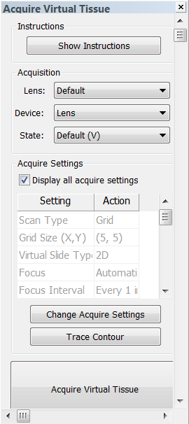 . The Virtual Tissue Acquire dialog box appears
  .
.Save the image in the desired location as prompted; name the image and select the file type from the drop-down menu:
![]() MBF recommends saving images as MBF JPEG2000 (either as .jp2 or .jpx). This file type retains additional information about the image unique to the system setup (pixel scaling, raw image data, application version, etc.). In the case of imaging issues, this information could serve as a diagnostic tool.
MBF recommends saving images as MBF JPEG2000 (either as .jp2 or .jpx). This file type retains additional information about the image unique to the system setup (pixel scaling, raw image data, application version, etc.). In the case of imaging issues, this information could serve as a diagnostic tool.
Grid: Use to define the area to be acquired as a set number of fields of view in a rectangular grid. This option requires that you be positioned at the top left corner of the region to be scanned; the current field of view is the first one scanned.
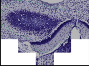
![]() Test the settings over a 3x3 grid before performing the full acquisition to ensure that the background image is satisfactory, that the trim and blend are appropriate, and to determine whether a stage delay is needed. This will save you considerable time!
Test the settings over a 3x3 grid before performing the full acquisition to ensure that the background image is satisfactory, that the trim and blend are appropriate, and to determine whether a stage delay is needed. This will save you considerable time!
Contour: Use a closed contour.

2D: Appropriate when only one image plane is desired.
3D: Appropriate when more than one image plane is needed to create an image stack. This is generally appropriate at higher magnifications, but can be used at lower magnification as well (3D is only available if you purchased the Virtual Tissue 3D module.)
Height of Virtual Tissue: Refers to the desired stack height. The values you choose will affect the processing time.
The number of planes in the stack is determined by the Stack Options.
![]() The program automatically adds the top plane to the number of focal planes.
The program automatically adds the top plane to the number of focal planes.
The total number of images to be acquired is dynamically displayed. This allows you to balance image data acquired with the time and disk space necessary to collect an image volume.
Manually, every: Check if it is necessary to manually focus the tissue
periodically throughout the scan. 
Automatically, every: This option requires special hardware. If this option is grayed out, the hardware is not installed or connected.
![]() Useful for stages that drift. Use manual focusing only if you need to repeat a completed scan that shows a significant loss of focus toward the bottom of the image to correct for drift.
Useful for stages that drift. Use manual focusing only if you need to repeat a completed scan that shows a significant loss of focus toward the bottom of the image to correct for drift.
If you are acquiring an image with brightfield illumination, the time delay is used to allow time for the stage in XY to stop moving and any vibration to die down.
For your test montage:
![]() This option has no effect on Virtual Tissue 3D acquisitions.
This option has no effect on Virtual Tissue 3D acquisitions.

Keep Image Open: Displays the image in the Image Organizer when the acquisition is complete for immediate inspection.
Multichannel Acquire: Uses the existing Multichannel acquire setup; adjust the Multichannel Acquire settings prior to performing a Virtual Tissue acquisition.
Compress Tile Files: Reduces the size of the image files.
Remove Temp Files: Individual image files are saved in a folder until they are compiled into the final image montage at the end of the acquisition.
Postpone Compilation: Individual image files will be acquired and saved in a designated folder, but a complete image will not be compiled automatically when the acquisition is finished.
Capture Virtual Tissue: Enter the distance in microns if the image needs to be acquired below the focus map.
![]() Useful if you want to create the focus map at the top of the tissue, but capture the image a set distance into the section thickness.
Useful if you want to create the focus map at the top of the tissue, but capture the image a set distance into the section thickness.

Pixel Trim Images: Removes rows and columns of pixels from the edges of each field of view (image tile).
This option allows you to correct for spherical aberration or a video card that acquires with a black or white strip on any of the image edges.
Try a test acquisition with all of these values set to zero, and increase them if there is a problem in the final image.
For low magnifications, 0-10 pixels is a suitable range.
For higher magnifications, larger trims (25-50 pixels) may be appropriate.
Seam Blending: Blends the edges from each field of view smoothly.
Background Color: Use if the image scan selected is Contour (only inside).
).