3. Preview
Cell detection workflow — Step 3
Purpose: This preview step is designed for fine-tuning the cell detection settings.
BrainMaker software displays the cell detection result for the current field of view in the software window.
You may want to zoom in to both speed up the preview process and to clearly see the results of cell detection with different settings applied.
Procedure
-
Select cell-detection settings in each section of the step 3. Preview window.
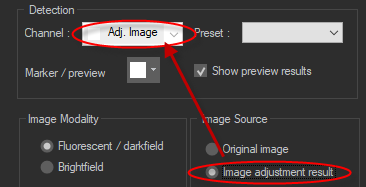
Detection setup
-
Channel: Select the appropriate color channel for cell detection depending on how the specimen is labeled.
Note that the channel is automatically set to Adj. image if you select Image adjustment result under Image Source.

- Preset: Click the drop-down to view and apply preset parameters for the cell detection if you have previously saved them. If there are no presets, you'll have the opportunity to save the parameters for this detection when you finalize the detection in Step 4.
- Marker/preview: Select a color that contrasts well with the image data. It will be used to mark cells in preview results and for the final cell-detection markers.
- Show preview results checkbox: Check or clear the box to view or hide the results of running the cell detection preview.
Image Modality: Select the image modality that most closely represents the type of image you are using.
Image source
Select the image-data you want to use for cell detection:
- Original image: Cell detection is based on the image as it appears onscreen.
- image Adjustment result: Modify the histogram in the Image Adjustment window to optimize the display for cell detection. If you select this option, the Channel near the top of the window is automatically set to Adj. image.
Cell Size
Set an approximate diameter for the largest and smallest cells to be detected. You can either:
- Enter values in the boxes corresponding to Large and Small diameter cells. OR
- Click the Measure button and follow the on-screen instructions to measure representative large and small cells using your cursor.
Detection
Choose detection using Cell Strength or Neural Network by clicking on the tab associated with your choice.
-
Cell Strength: based on signal strength in relation to background signal.
Use the slider or type a number into the box to specify the filter strength; the higher the value, the more background noise is removed.
-
Neural Network: Uses machine learning in conjunction with neural-network classifiers to identify neuronal cell subtypes.
-
Use the slider or type a number into the box to specify the filter strength.
-
Select a neural network from the drop-down menu.
-
-
-
Click Preview and review the cell detection results. Adjust settings as needed and repeat.
-
When you are satisfied with the preview results, click Next Step to move to the final step of the workflow, step 4. Filter and Finalize.
Commands present in all steps of the workflow
 Previous step / Next Step: Click to advance in the workflow or revisit a previous step. Alternatively, you can click the steps listed at the top of the workflow to jump to that step.
 New workflow: Click the new workflow button to start over; the settings will revert to those specified in the previous completed workflow.
 Presets: Click to select a preset and apply those saved settings to the current workflow. Note that you can save your settings as a preset at the end of the workflow.
- Loading an existing preset: Click Presets and select a preset from the list.
-
Saving or deleting presets: Click Presets and select Edit Presets; the Save/Update dialog box opens. You can:
- Type a name for the preset, and click Save.
- Select an existing preset and click Delete to remove it.