Detecting spines (3D)
See tutorial: Detect, classify and edit spines (3D)
Editing spines (3D), Analyzing spines and synapses with Neurolucida Explorer
You need to trace the trees before you can detect spines. To display the Detect Spines panel, click the Spine button.
Detecting spines
- Click Detect All for an initial detection with the default settings. Automatic detection may take a few seconds (see How are spines detected? below to learn about the detection process).
The program renders the spines that meet the criteria defined below using a surface mesh. Because rendering is based on a different algorithm than the one used for spine detection, it might not reflect exactly the spine profiles generated by the spine detection process.
- Inspect the results of the detection and adjust the detection settings before detecting spines again.
- Outer range: Maximum distance between the dendritic surface and the potential top of the spine head.
- Minimum height: Minimum distance between the dendritic surface and the furthest voxel identified in the spine.
- (Detector sensitivity: Use when detecting individual spines.)
- Minimum count: Adjust to ignore objects that are too small to be considered spines. Works in conjunction with Minimum height.
- Optional: To detect spines in addition to spines already detected with Detect All, check the Keep existing spines box.
- Click Detect All to detect again.
This is useful for testing the spine detection settings on a small region of a large image as it restricts automatic detection to the region near the branch.
- Under Detection, adjust the settings.
- Check the box Click image to detect all on nearest branch.
- Click near the desired branch to detect all the spines on that branch automatically.
- Zoom, rotate, and pan as needed to view the spine of interest clearly.
- Adjust Outer range and Detection sensitivity as necessary. By clicking to detect a spine, you are overriding the Minimum height and Minimum count settings.
- Outer range: Adjust the maximum distance between the surface of the tree and the potential top of the spine head.
- Detector sensitivity: Adjust to identify spines that might be too light or too dark based on the original image intensity.
- Click the spine of interest. The detected spine is displayed with a color overlay.
You may see a warning: "Unable to detect a spine at the selected location." Under Detection Settings, increase the Detector sensitivity (e.g., from 100% to 125%) then click the spine again to detect it.
- If you can't identify parameters that produce good results for all the spines, run the automatic detection several times (each time with different parameters) with the Keep existing spines box checked.
- After each automatic detection, delete the detected spines that were not correctly modeled: Click the Edit button to switch to Edit mode, click a spine to select it, press the Delete key, then click the Edit button again to return to Detection mode.
- Keep existing spines is used to ensure that the spines modeled properly with one set of parameters are not deleted or modified by the next automatic detection.
- If you notice that a few spines remain undetected, use the “click to detect” type of modeling (i.e., modify Outer range and /or Detector sensitivity then click the spine to model it).
- For a large complex cell with many branches, detect on a single branch first to parameterize more quickly: check the Click to detect all spines on nearest branch box and click the desired branch.
- If the modeled spine head appears larger than the actual spine head, reduce the detector sensitivity.
- If the modeled spine neck spreads out onto the dendritic shaft, increase the surface clearance.
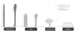
Occasionally, a spine might be represented as stick display. This is because the spine is too small for the software to create the 3D overlay. 
Deleting or moving points may lead to deleting the detected spines potentially associated with these points. Make sure to detect the deleted spines again.
Optional
- If your image is “noisy,” check the Filter image noise box.
- To detect spines in addition to spines previously detected manually, check the Keep existing spines box.
Classifying spines
Once the spines have been detected (automatically or manually), click Classify All. Spines are classified according to four types (filopodium, mushroom, stubby, thin) and color-coded based on the default settings.
- To change the classification default settings, click the Settings button.
The spine types are determined from a combination of measured values and derived ratios that you can modify here.
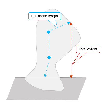
- By default, the program uses the spine backbone length for classification, but you can select the spine total extent instead by checking the corresponding box.

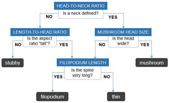
- The flowchart illustrates how the program uses the settings you define to assign one of four types to each detected spine:

- The measurements are illustrated here:

- Optional: To classify additional spines without changing the current classification of spines already detected, check the Keep existing classification box.
- To re-classify manually: Click the Edit button, click a spine to select it, and select a different type from the Type drop-down menu in the panel.
How are spines detected?
Once you've clicked the detect All button or clicked individual spines to detect them, the program takes a series of steps.
- The dendritic branch is refined and displayed with an axial smear correction.
- The program uses image segmentation to identify all the foreground voxels that are outside of the volume occupied by the dendritic model.
- The identified voxels are clustered based on image intensity and distance to the dendritic model (defined in the Detect Spines panel). Clustering takes into account the adjacent spines scenario and leads to the identification of spine candidates.
- Only the spines candidates that meet the detection criteria are rendered using a surface mesh (based on the Marching Cubes algorithm). The mesh is further processed to remove surface irregularities.
- Spine extent >= Minimum height
- Voxel count >= Minimum count
- Ratio spine extent/base>= 0.4
- Spine extent<=Outer range
- The spine models are displayed using multiple colors to help discriminate between adjacent spines. The spines have not been classified.
To change the color of individual spines, and split, merge or delete spines, display the Edit Spines panel.
A spine is detected and rendered if it meets all these criteria:
The spine extent is based on image data; it is the shortest distance from the furthest identified voxel to the surface of the dendrite. It does not necessarily follow the spine neck to the dendritic surface.
The minimum height can be defined in the Detect Spines panel.
The voxel count is based on voxel processing and is used to evaluate spine size. It is determined by the image scaling.
The Minimum count can be defined in the Detect Spines panel.
The spine extent is based on image data; it is the shortest distance from the furthest identified voxel to the surface of the dendrite.
The base is determined using the Rayburst diameters calculated for spine detection. It is an evaluation of the spread of the spine at the surface of the dendrite and it takes into account axial smear correction.
The outer range is the maximum distance from the dendritic surface to the potential top of the spine head. It can be defined in the Detect Spines panel.
Renderings might not reflect exactly the spine profiles generated by the spine detection process since detection and rendering use different algorithms.
How does the automatic spine classification process work?
Once you've clicked the Classify All button, the program processes the detected spines.
- Spines are classified into canonical types (stubby, mushroom, thin, or filopodium) based on the method described in Automated Three-Dimensional Detection and Shape Classification of Dendritic Spines from Fluorescence Microscopy Images (Rodriguez, Ehlenberger, Dickstein, Hof, & Wearne, 2008).
- Color-coding is applied to represent spine classification. Each spine classification has been assigned a color using Spine preferences.
Reference
Dickstein, D.L., Dickstein, D.R., Janssen, W.G.M., Hof, P.R., Glaser, J.R., Rodriguez, A., O'Connor, N., Angstman, P., and Tappan, S.J. (2016). Automatic dendritic spine quantification from confocal data with Neurolucida 360. Curr. Protoc. Neurosci. 77:1.27.1-1.27.21. doi: 10.1002/cpns.16
Rodriguez, A., Ehlenberger, D.B., Dickstein, D.L., Hof, P.R., and Wearne, S.L.. (2008). Automated Three-Dimensional Detection and Shape Classification of Dendritic Spines from Fluorescence Microscopy Images. PLoS ONE, 3(4), e1997. http://doi.org/10.1371/journal.pone.0001997