Preparing for tracing
Tracing a live image
Follow these steps to display and move around the specimen displayed in the Neurolucida tracing window.
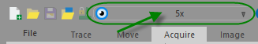
- If you have a motorized system, go to the Acquire ribbon and select the camera from Device controls > Devices.
- Click Acquire>Display>Live Image to display the tissue on your screen. You may also need to adjust the light levels directly on your microscope.
- If your system is not motorized, select an objective and the corresponding software lens.
- Rotate the microscope's nose piece to select the appropriate objective.
Select the corresponding lens from the Lens drop-down menu.
Lenses must be correctly calibrated. See Calibration overview to learn how to set up and check calibration.
- Prepare your workspace. From the Workspace ribbon, open the most commonly used tools: Macro View , Z meter, Camera histogram, Camera settings and Stack acquisition.
- Choose a location for the reference point. The reference point is essential to maintain alignment between tissue and traced reconstructions.
- Manual stage: Move the stage until the desired location is within the field of view.
- Motorized stage: Use the joystick to move to the desired location.
- Click to place the reference point.
-
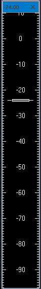
Verify that the program is registering movement along the Z-axis.
As you focus up and down using the focus knob on the joystick (or the focus on the microscope if equipped with an internal Z motor), verify that Z is changes accordingly in the Z meter and that you move through planes on the screen.
- You're ready to start tracing. Go to the Trace ribbon to view the tracing functions.
Tracing an acquired image
- Open the image or image stack.
-
If the image was acquired with an MBF Bioscience system, go to step 3; if not, verify that the scaling is correct—this is critical!
Correct scaling means that the program uses the same scaling (i.e., micron-to-pixel or micron-to-voxel ratio) as the scaling that was used when the image was acquired.
-
If scaling is embedded in the image, the program applies the correct scaling. If there is no software lens associated with this scaling, you are prompted to define a lens so that the image is displayed at its real/original size.
-
If no embedded scaling is identified, do one of the following:
- Provide or override the scaling
- Image an artifact of a known length and use it to create a new lens.
- Measure an artifact of a known length with Trace>Measure line to verify the scaling.
-
-
Verify that Neurolucida software is registering movement along the Z-axis.
-
Display the Z Meter (Workspace>Views>Z meter).
-
Focus up and down using the mouse wheel or the Page Up/Page Down keys.
-
- You're ready to start tracing. Go to the Trace ribbon to view the tracing functions.
See also Tracing Trees in Single Sections, Tracing Trees in Serial Sections, Tracing Contours, Serial Section Reconstruction