MBF Bioscience Blog

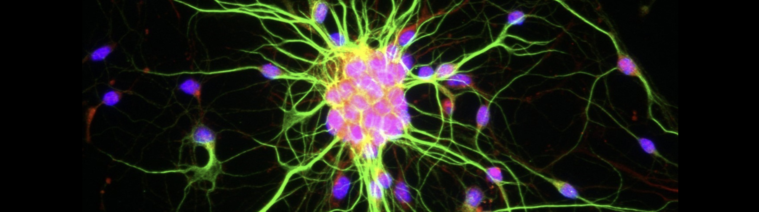
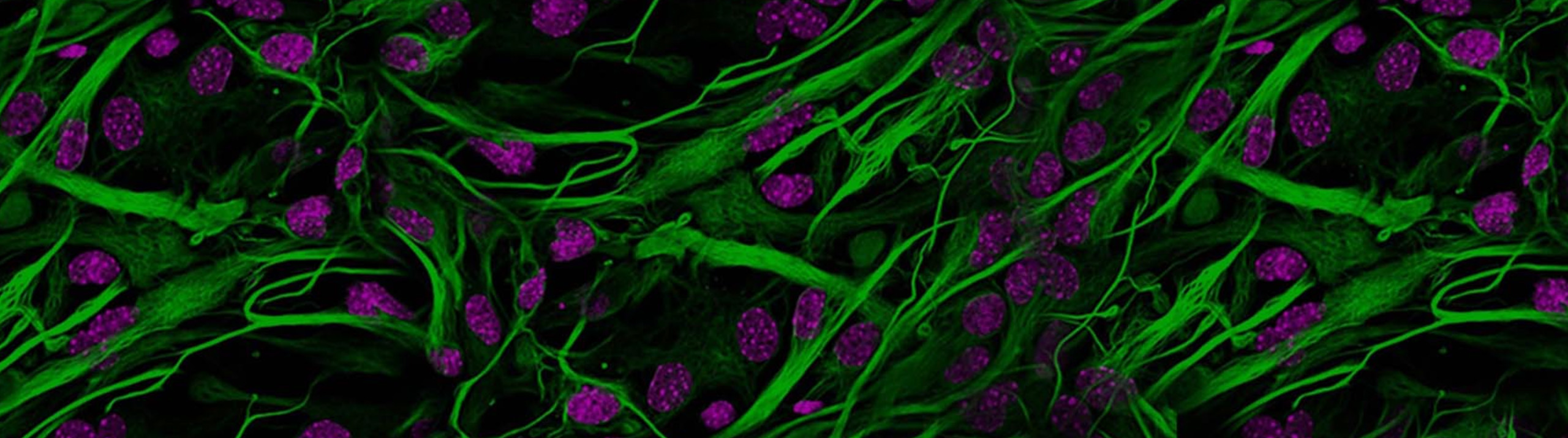
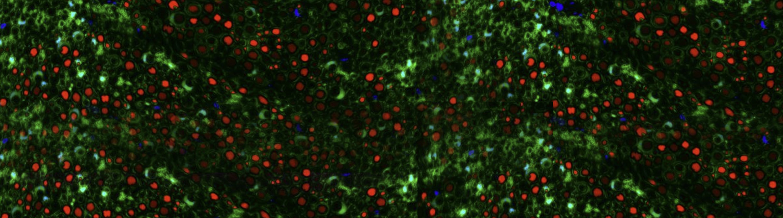
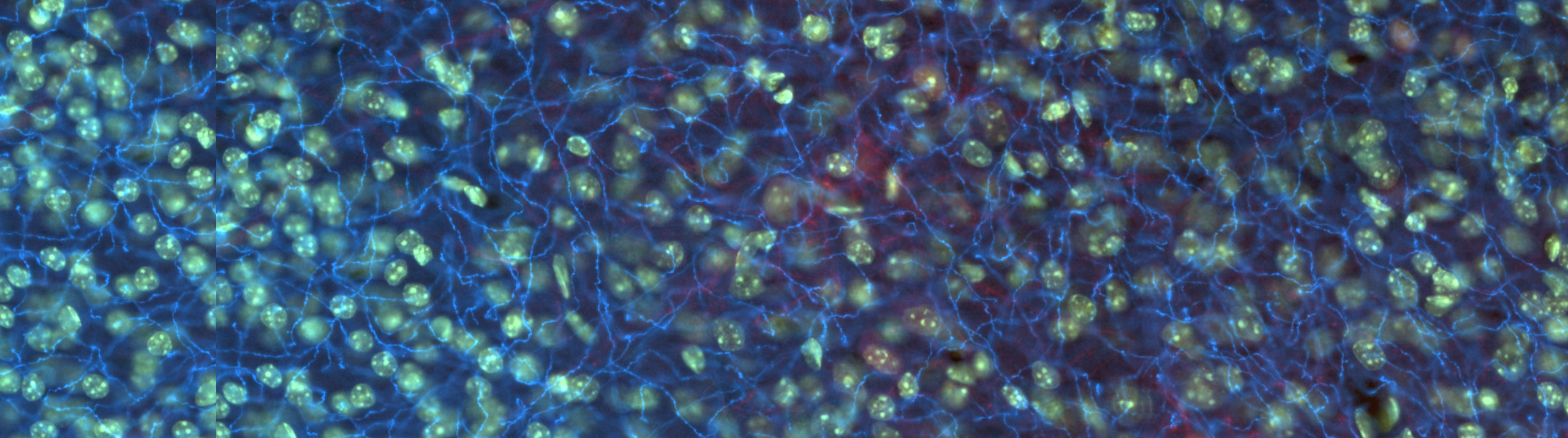
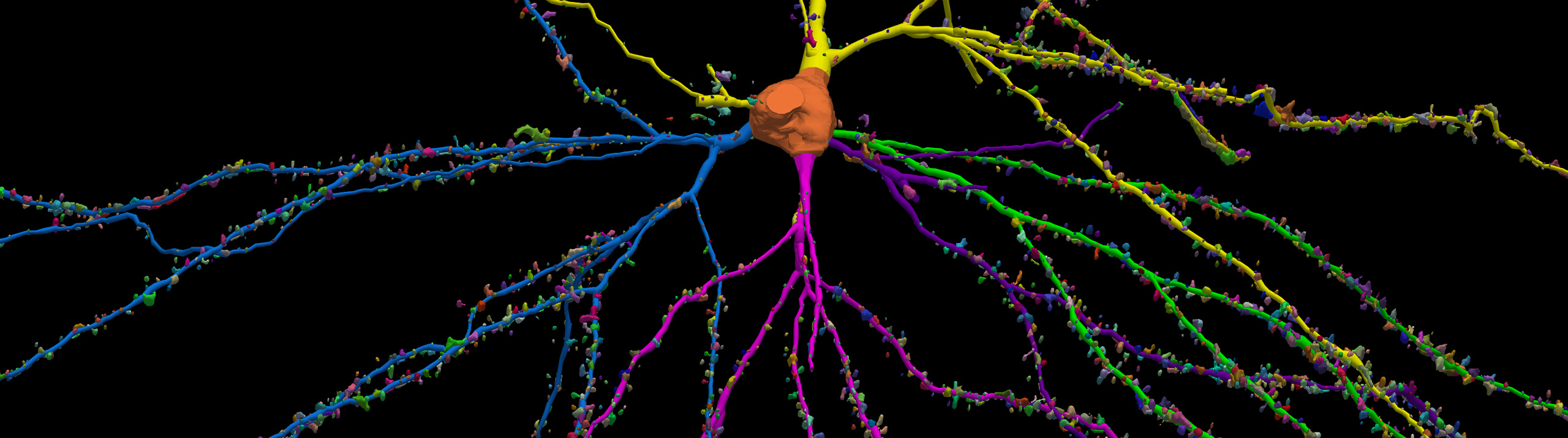
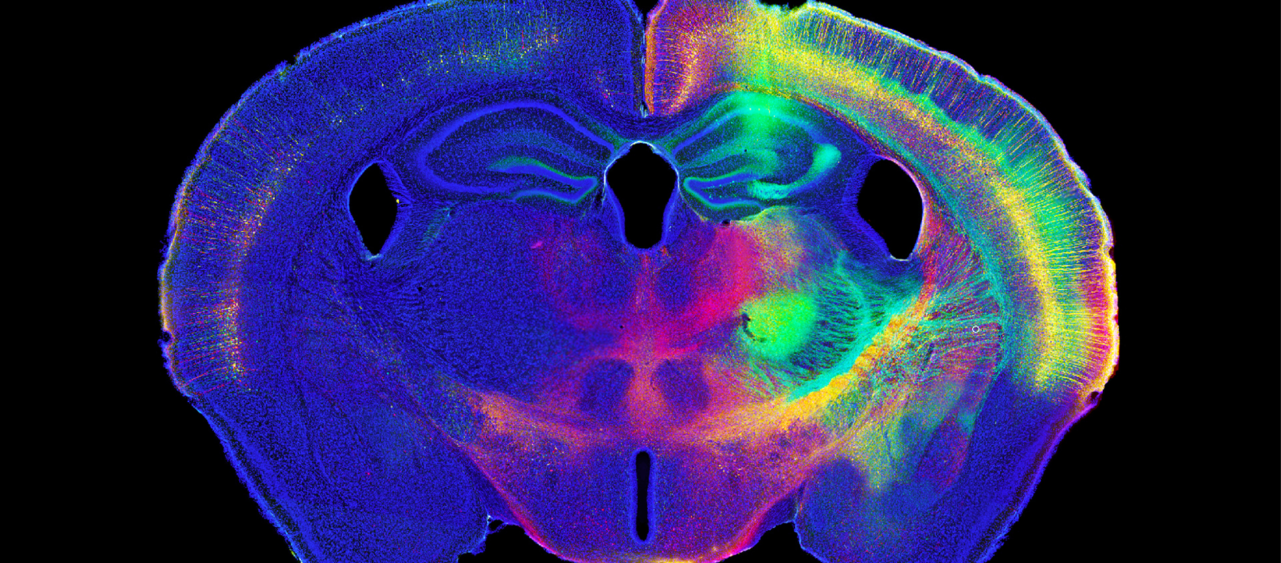
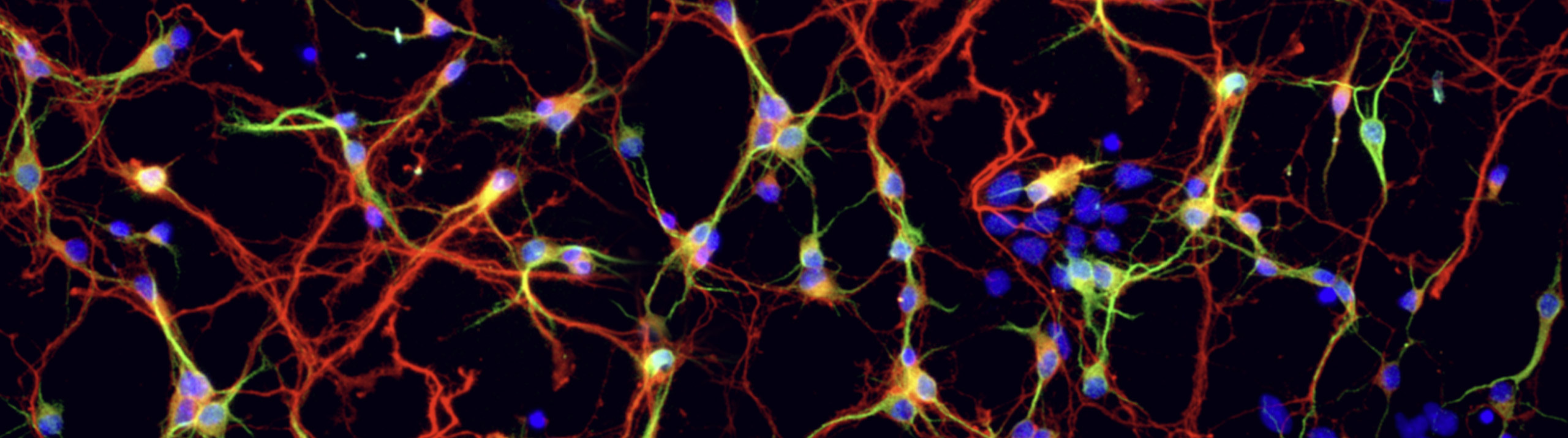
Diversity of Enteric Glial Cells - April NeuroArt Juror's Choice Winner Marissa Puzan, Boston University MBF Bioscience and the Journal of Neuroscience Research announced today that beginning this month, the Juror’s Choice winner in the NeuroArt image contest will be featured on the cover of the Journal of Neuroscience Research. Winners also have the opportunity to publish an editorial article or a research paper in the journal....

Read More

Neuroscientists can now analyze the size and complexity of neurons and collect unbiased stereology data with greater speed and efficiency We are happy to announce the release of Neurolucida and Stereo Investigator version 2017. This version features a completely revamped user interface that’s intuitive and easy to navigate.   “Neurolucida and Stereo Investigator version 2017 are completely redesigned to improve the user experience and increase productivity,” said Jack...

Read More

Following a well-designed protocol is essential to achieving accurate and consistent results in scientific research. Now, scientists using Neurolucida 360 for dendritic spine and neuron analysis can follow a published set of guidelines to ensure optimal confocal data series for proper dendritic spine quantification and neuron reconstruction. The paper, written by MBF Bioscience scientists and researchers from the Icahn School of Medicine at Mount Sinai...

Read More

Astrocytes (GFAP) in the dentate gyrus of a mouse hippocampus. Image courtesy of Dr. Ahmad Salehi, Stanford University.   It is well known that physical exercise eases the symptoms of neurodegenerative disorders like Alzheimer’s disease and helps to prevent their onset. Researchers at Stanford University are working on figuring out how it happens.   In their study, published in the journal Brain Structure and Function, scientists in Dr....

Read More

MBF Bioscience is sponsoring a new image contest that encourages scientists and artists to share their views of the brain   Williston, VT— The NeuroArt image contest brings together scientists, artists, and neuroscience enthusiasts from around the world to share their view of the brain. Any image of the brain is accepted, including but not limited to microscope images, pencil drawings, and paintings.   Eligible contestants can visit neuroart.com/image-contest...

Read More

[caption id="attachment_6526" align="aligncenter" width="800"] Representative images of Iba-1+ microglia in the postnatal day 10 rat hippocampus. Image courtesy of Anna Klintsova, PhD.[/caption] Children born with fetal alcohol spectrum disorders face a range of physical and cognitive impairments including long-term deficits in learning, behavior, and immune function. In a paper published in Neuroscience, Dr. Anna Klintsova and her lab at the University of Delaware report that activation...

Read More

Complex imaging and experimental tasks must happen quickly in a smooth workflow so that experiments can be performed within the time constraints imposed by working with behaving animals. ScanImage is the software of choice for controlling your scanning microscopy experiments because it provides robust cutting edge imaging features coupled with the ability to automate and time workflows like no other application can. A key feature...

Read More

The optical fractionator probe was used to quantify the number of neurons and glia in the dentate gyrus   Doctors have used lithium to treat patients with bipolar disorder since the 1970s. Known for its efficacy in stabilizing patients' moods by regulating manic episodes, lithium is also associated with a decreased risk of suicide. But while this naturally occurring element is the most widely prescribed medication for...

Read More

[caption id="attachment_6403" align="aligncenter" width="632"] This figure illustrates the separate and combined effects of acute stress and fear conditioning/extinction on dendritic morphology of pyramidal neurons in the infralimbic region of medial prefrontal cortex. Each neuron shown is a composite made up of apical (blue) and basilar (orange) arbor near the mean of the group. The apical and basilar arbors of each composite are from different neurons....

Read More

The 3 algorithms in Neurolucida 360 were used in combination to create a smooth, accurate reconstruction   Minor releases typically don't include new features, but Neurolucida 360 isn't an ordinary piece of software. Neurolucida 360 v2.7 has many new features and improvements, including: A new automatic tracing algorithm - Rayburst Crawl Capture videos of your rotating neuron reconstructions for presentations and publications New backbone length analysis for dendritic spines Improved handling of images...

Read More