Iron Deficiency Worsens Fetal Alcohol Spectrum Disorders
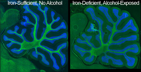
An immunostained image of myelin basic protein in the cerebella of a mouse brain with an iron-sufficient diet compared with the brain of a mouse exposed to alcohol and fed an iron-insufficient diet. It shows the reduced cerebellar size due to the ID-alcohol combination. Green is MBP immunostain, blue is DAPI for nuclei. Image courtesy of Susan Smith, PhD.
If a pregnant woman drinks alcohol, she risks giving birth to a baby with physical and cognitive deficits – characteristics of fetal alcohol spectrum disorders. In a new study, researchers say that when the mother is low in iron, the consequences are even worse.
The scientists examined two groups of pregnant rats – one group was fed an iron sufficient diet while the other was fed a diet with insufficient iron levels. The offspring from both groups were exposed to alcohol from 4 to 9 days after birth – a time when their brains are going through a growth spurt and are particularly sensitive to alcohol. They were compared to offspring who received an iron-sufficient diet but were not exposed to alcohol. This growth spurt correlates to a growth spurt in humans that occurs during the third trimester of pregnancy.
The researchers used delay and trace eye blink classical conditioning methods to assess the offspring’s learning and memory. Learning impairments were reported in both alcohol-exposed groups regardless of their iron status, but more extreme impairments were seen in iron deficient rats compared to iron sufficient rats. After the behavioral tests were completed, the researchers studied the cerebellum and hippocampus – brain regions involved in learning and memory – at a cellular level.
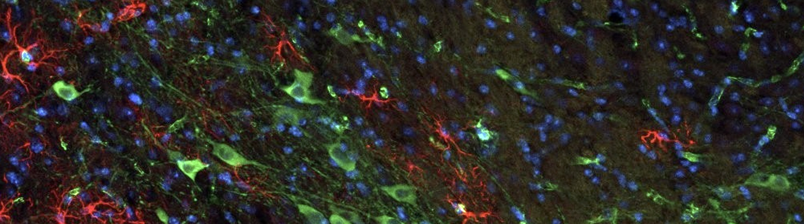
Using the Optical Fractionator probe in Stereo Investigator, the research team quantified neurons in two different areas of the rat brain: the cerebellar interpositus nucleus and the CA1 region of the hippocampus. Their unbiased stereological analysis revealed significant neuronal loss in the alcohol-exposed iron deficient rat brains compared to the iron sufficient rat brains.
“The most important finding from this study is that maternal iron status strongly influences alcohol’s neurobehavioral damage in the developing offspring,” the authors say in their paper. These data endorse that the treatment of maternal [iron deficiency] through normalization of iron status should improve the child’s developmental outcome despite the gestational alcohol exposure.” (Huebner, et al.)
Huebner, S.M., Tran, T.D., Rufer, E.S., Crump P.M., Smith S.M., (2015) Maternal iron deficiency worsens the associative learning deficits and hippocampal and cerebellar losses in a rat model of fetal alcohol spectrum disorders. Alcoholism: Clinical and Experimental Research doi: 10.1111/acer.12876.