
Hippocampal Neurons Change After Melatonin Injection
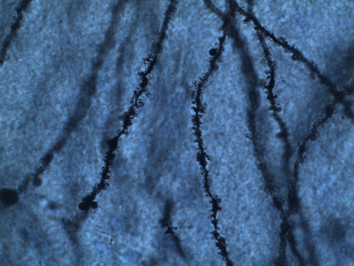
Representative dendrites of dentate gyrus neurons of Siberian hamsters injected with melatonin (stained with Cresyl violet). Ikeno et al found hamsters injected with melatonin displayed decreased spine density on neurons in the dentate gyrus. Image courtesy of Tomoko Ikeno, Ph.D.
Night falls and a powerful hormone called melatonin kicks in. The gears of the circadian clock are turning as you get ready for bed and soon drift off to dreamland. But all is not quiet in the brain. In response to the circadian rhythm, neurons are transforming.
A new study published in the journal Hippocampus found that melatonin prompts dendrites to grow longer in one part of the brain, while in another part the hormone causes dendritic spine loss.
In their study, scientists at Ohio State University injected Siberian hamsters with a dose of melatonin in the afternoon, several hours before a natural increase in the hormone would normally occur. Four hours after the injection, they used Neurolucida to examine sections of their brains, reconstructing neurons in two areas of the hippocampus – the CA1 and dentate gyrus. They then used the software to calculate the number of branch points and length of dendrites in their reconstructions. What they saw was longer, more complex dendrites in the CA1 region of the hippocampus of hamsters that received melatonin versus those that received a placebo. Then they analyzed spine density, finding that hamsters that received melatonin had decreased spine density in the dentate gyrus than the control group.
“By using Neurolucida, we found that melatonin treatment induced rapid remodeling of hippocampal neurons and induced a nighttime state of the hippocampal neuronal morphology,” said Dr. Tomoko Ikeno, who worked with Dr. Randy Nelson on the study.
The “nighttime state” she refers to is characterized by the presence of certain hormones produced during the dark hours of night. In their analysis, the researchers saw elevated levels of Period1 and Bmal1 after melatonin injection. These hormones are expressed by genes associated with the circadian clock, and their presence offers evidence that “melatonin functions as a nighttime signal to coordinate the diurnal rhythm” and that this rhythm compels hippocampal neurons to change structurally, according to the paper.
Ikeno, T. and Nelson, R. J. (2014), Acute melatonin treatment alters dendritic morphology and circadian clock gene expression in the hippocampus of Siberian Hamsters. Hippocampus. doi: 10.1002/hipo.22358