Dendritic Spine Loss Reported in Schizophrenia and Bipolar Disorder
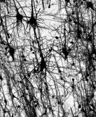
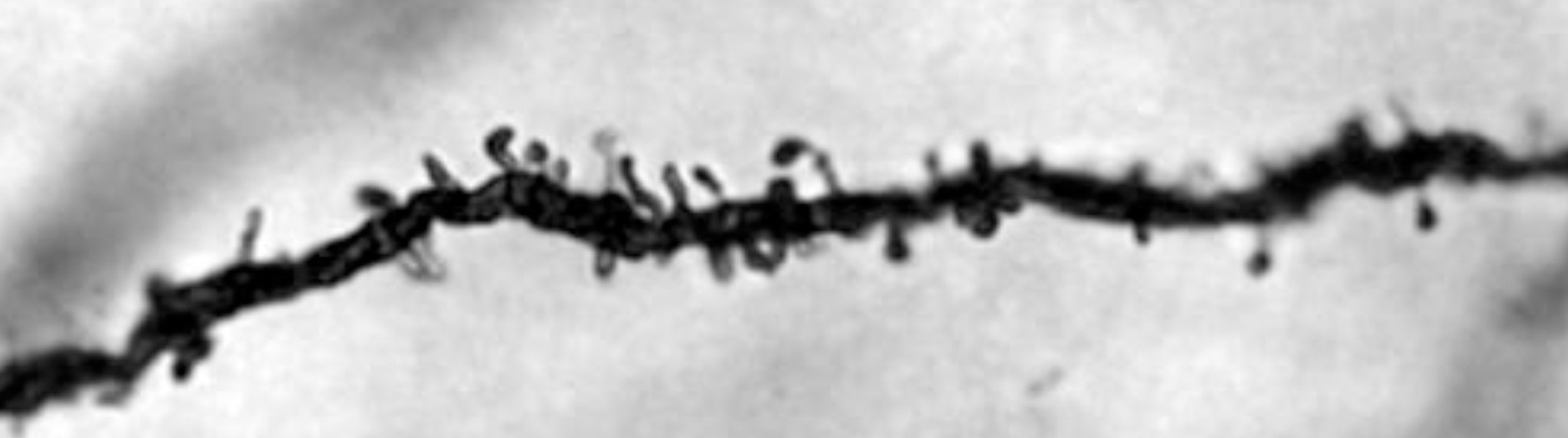
Golgi-stained human brain tissue from the dorsolateral prefrontal cortex.
Schizophrenia and bipolar disorder are very different mental illnesses, but researchers are discovering evidence that the two disorders have some common pathologies. According to a recent study, a shared characteristic appears to be dendritic spine loss.
The researchers used Neurolucida to study pyramidal cells in human brain tissue from individuals with schizophrenia (n=14), individuals with bipolar disorder (n=9) and unaffected control participants (n=19). The pyramidal cells were located in the dorsolateral prefrontal cortex – a region that plays a key role in working memory. Bipolar patients showed significantly reduced spine density (10.5 percent) compared to control. Schizophrenic patients also showed lower spine density (6.5 percent), but this number just missed significance when compared to control patients. Individuals with both illnesses showed a lower number of spines per dendrite, as well as reduced dendritic length compared to controls.
To obtain these results, researchers analyzed 15 Golgi-stained pyramidal cells in each tissue sample. They used Neurolucida to reconstruct the longest dendrite on the pyramidal cells and to mark spines. After the researchers finished reconstructing the cells, Neurolucida provided them with important data about the dendrites and spines.
“It would have been very difficult, if not impossible, to obtain the measurements for this study without Neurolucida,” said lead investigator Dr. Glenn Konopaske. “Other programs I am aware of allow measurements of dendritic spine density along only a portion of the dendrite. Neurolucida allowed an assessment of spine density along the whole dendritic branch.”
Dr. Konopaske’s research confirms previous studies, which have shown spine loss on pyramidal cells in the dorsolateral prefrontal cortices of individuals with schizophrenia, but according to the paper, the level of reduction is lower (6.5 percent) than previously reported (23 percent) – a finding, which the authors say suggests “variability in spine pathology in SZ.”
“The current study suggests that spine pathology is common to both SZ and BP. Moreover, the study of the mechanisms underlying the spine pathology might reveal additional similarities and differences between the two disorders, which could lead to the development of novel biomarkers and therapeutics,” the authors say.
Konopaske, G.T., Lange, N., Coyle, J.T., Benes, F.M. (2014). Prefrontal Cortical Dendritic Spine Pathology in Schizophrenia and Bipolar Disorder. JAMA Psychiatry. doi: 10.1001/jamapsychiatry.2014.1582.