Hawaii Scientists Measure Density of Parvalbumin-Interneurons With Stereo Investigator
Foods like tuna fish and Brazil nuts are rich in selenium, a mineral that scientists say has antioxidant effects, keeping the brain healthy and free of clutter so cells can work smoothly together. A key element of this process is Selenoprotein P (Sepp1) – a protein that delivers selenium to neurons by binding with another protein – ApoER2. Neuroscientists at the University of Hawaii say Sepp1 plays a critical role in brain function, and deficits may play a part in mental illnesses like schizophrenia.
In their study published in Neuroscience, the researchers investigate the relationship between Sepp1 and parvalbumin (PV)-interneurons – a class of brain cell that controls firing rates and synchronizes spiking activity among other groups of neurons. Previous research shows that these cells need selenium to develop properly, so the scientists set out to find out what affect a Sepp1 deficit would have on the mouse brain.
Led by Dr. Matthew W. Pitts, the research team compared the brains of wild type mice with Sepp1 deficient mice. They used a Zeiss Axioskop microscope equipped with Stereo Investigator to conduct a stereological analysis of PV-interneurons in several different regions of the mouse brain. Using Stereo Investigator’s optical fractionator probe, they observed reduced numbers of PV-interneurons along with elevated oxidative stress in the inferior colliculus of Sepp1 deficient mice, a region involved in processing auditory information.
“Stereo Investigator was particularly useful for estimating cell density in larger brain structures, such as the inferior colliculus,” said Dr. Pitts.
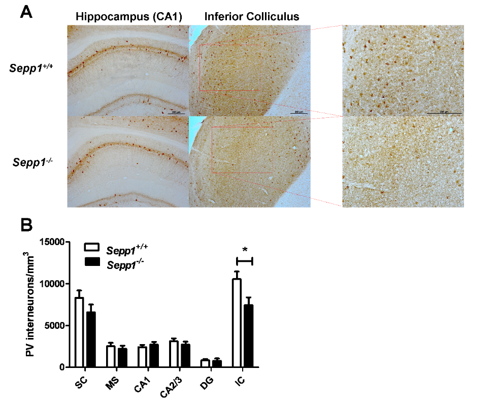
Reduced density of PV-interneurons in Sepp1-/- mice. (A) Representative images showing PV expression in the hippocampus (left column) and inferior colliculus (middle and right columns) of WT Sepp1+/+ (top row) and Sepp1-/- (bottom row) mice. Higher magnification images of the inferior colliculus (far right) (B), Mean density of PV-interneurons per mm3 (+-SEM, n=6 per genotype) in brain regions investigated: SC; MS; DG, CA1, and CA2/3 of the hippocampus; IC. * P<0.01. Figure courtesy of Matthew W. Pitts, Ph.D.
Since scientists speculate that dysfunctional PV-interneuron networks may be involved in neuropsychiatric conditions, the researchers conducted behavioral tests that showed impairments in contextual fear extinction, latent inhibition, and sensorimotor gating in the Sepp1 deficient mice – behaviors observed in some mental illnesses.
“Previous studies (Valentine et al., 2008) and our findings together indicate that ApoER2- mediated uptake of Sepp1 serves an important neuroprotective role in the inferior colliculus,” the authors say in their paper. “These findings may have relevance to neuropsychiatric conditions in which dysfunc- tional PV-interneuron networks have been implicated, such as epilepsy and schizophrenia.”
Pitts M.W., Raman A.V., Hashimoto A.C., Todorovic C., Nichols R.A., Berry M.J. Deletion of selenoprotein P results in impaired function of parvalbumin interneurons and alterations in fear learning and sensorimotor gating. Neuroscience. 2012 Apr 19;208:58-68. doi: 10.1016/j.neuroscience.2012.02.017.