Stereo Investigator Helps Scientists Assess Damage in Rat Model of Ischemic Stroke
A stroke patient is rushed to the hospital. Deprived of oxygen-rich blood, brain cells have already died, and more damage will probably occur in the hours and days to come. But researchers at the University of South Florida and the University of Padova in Italy say a two-part package administered through the body, rather than directly into the brain, may be the key to staving off some of the cell death that takes place after a stroke.
In their study, published in the Journal of Enzyme Inhibition and Medicinal Chemistry, the scientists saw a smaller region of damage in a rat model of focal cerebral ischemia, when the rats were treated with a combination of an anesthetic and a Caspase-3 inhibitor – a drug that suppresses a protein involved in brain cell death.
According to the study, Caspase inhibitors are normally injected directly into the brain, diverting the need to pass through the protective blood-brain-barrier. But the authors, relying on evidence from previous studies that describe a disruption in the blood-brain-barrier after cerebral ischemia, found that Caspase-3 inhibitors were able to reach their cerebral targets after being injected into the body. “BBB disruption allows the [intraperitoneal] administration of caspase-3 inhibitor in order to assess perinfarct tissues, circumventing the need for invasive intracranial administration,” the authors say in their paper.
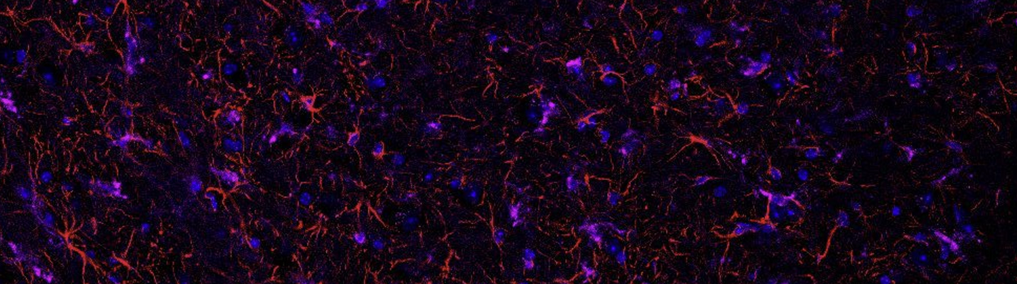
The researchers neurologically evaluated the rats based on motor function at days zero, four, and fourteen post ischemia. At the two-week mark, they euthanized the animals, sectioned their brains, and used Stereo Investigator to carry out an unbiased stereological analysis of the damaged sites. They quantified damaged cells with the optical fractionator and optical disector probe and used the Cavalieri estimator to measure the volume of the damaged region.
“In the neurological score, we found that the combination of anesthetics with caspase inhibitors substantially improve neurological deficit 4 days after the insult,” the authors say in their paper. “Our results also suggest that intraperitoneal injection of caspase-3 inhibitor in combination with Isoflurane or Propofol has a significant effect in reducing the volume of the infarct, when the injury was assessed at 14 days following the insult. However, tunel-positive cells and cleaved caspase-3-positive cells numbers were significantly decreased by the anesthetic-caspase inhibitor combination or the caspase-inhibitor alone, but not by the anesthetics alone.”
[Chaparro, E., Erasso, D., Quiroga, C., Bosco, G., Parmagnani, A., Rubini, A., Mangar, D., Camporesi, E. (2012). Repetitive intraperitoneal caspase-3 inhibitor and anesthesia reduces neuronal damage. Journal of Enzyme Inhibition and Medicinal Chemistry, (00), 1-7.
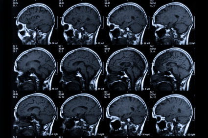
Image: MRI scan of human head via iStock Photo.