UVA Scientists Use Neurolucida in Study Identifying Two New Circuits in Rat Neocortex
Revving engines, blasting sirens, the drummer next door. Despite the myriad sensory stimuli going on around us at any given moment, humans have the ability to stay focused on the task at hand. This skill is due to a part of the brain known as the neocortex, a six-layer structure whose intricate wiring is largely a mystery. But researchers at the University of Virginia just took a big step toward a broader understanding of how this region works. They discovered two never-before-identified circuits in the rat sensorimotor cortex that help explain how the brain filters information.
“We identified two previously unknown and distinct cortical interneuronal circuits that link input-receiving L1 interneurons via L2/3 interneurons to output-producing L5 pyramidal neurons in the rat sensorimotor cortex,” the authors say in their paper published January 13 in Nature Neuroscience.
One circuit links single-bouquet cells (SBC), a type of neuron found in the first level (L1) of the neocortex to seven different types of interneurons found in levels two and three (L2/L3), which connect to pyramidal neurons in level five (L5). In the second circuit, L1 elongated neurogliaform cells (ENGCs) link to L5 pyramidal neurons via three types of L2/L3 interneurons. The two circuits work together to filter out “noise” and allow for a concentrated focus on important information, the study says.
To achieve this discovery, the researchers used the patch clamp electrophysiology technique to record the morphology of up to eight neurons at a time. They tested “14,832 connections between 1,703 L1 neurons, 3,120 L2/3 interneurons and/or 3,394 L5 pyramidal neurons in the cortical slices.”
Instead of using firing patterns to identify L1 SBCs, as had been done in previous studies, they classified the cells based on the arborization patterns of their axons, which differed from those of ENGCs. They also found that SBCs and ENGCs connect to L5 pyramidal neurons at different points, regulating their behavior in different ways.
“These results suggest that SBC → L2/3 interneuron → L5 pyramidal neuronal circuits are structured to disinhibit a small population of L5 pyramidal neurons, whereas ENGC → L2/3 interneuron → L5 pyramidal neuronal circuits are organized to inhibit a large population of L5 pyramidal neurons,” the authors say in their paper.
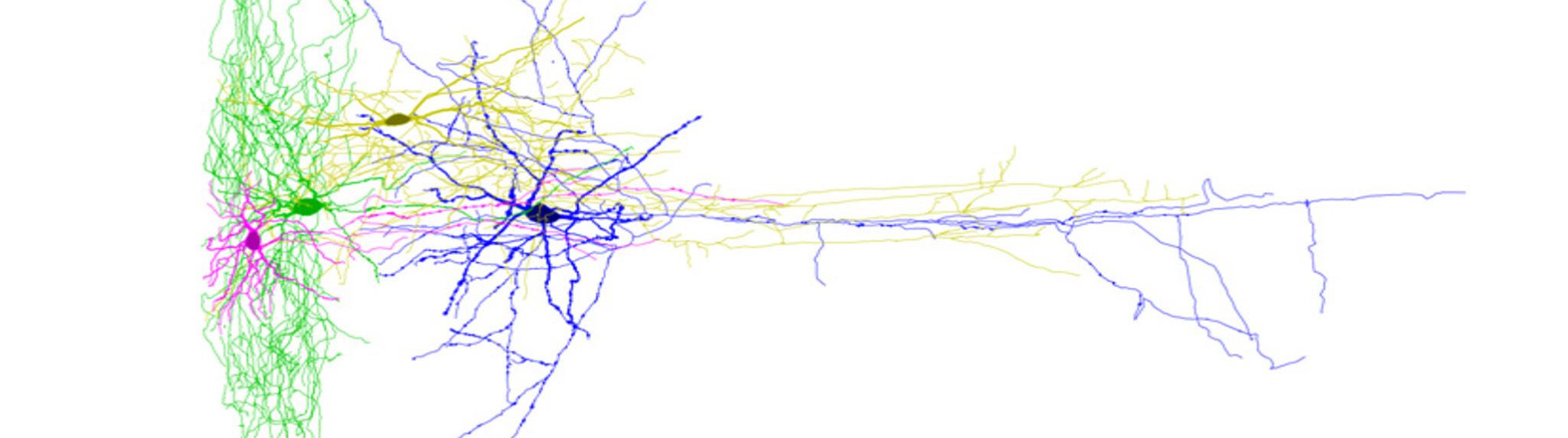
They used Neurolucida to reconstruct and quantitatively analyze the morphologically recovered neurons. “Neurolucida allowed for accurate neuronal reconstruction in order to do precise measurements on the axonal and dendritic lengths, branching, and contacts essential to the analysis necessary for the major findings in this work,” said Dr. Ruth Stornetta, an author of the study. “The tracing software is intuitive, easy to learn, and a joy to use,” she added.
Jiang, X., Wang, G., Lee, A. J., Stornetta, R. L., & Zhu, J. J. (2013). The organization of two new cortical interneuronal circuits. Nature neuroscience,16(2), 210-218. doi:10.1038/nn.3305.